Thouvenot et al. Ipsilateral uveitis and optic neuritis in multiple sclerosis.Mult Scler Int. 2012;2012:372361. doi: 10.1155/2012/372361
Background: Uveitis is 20 times more frequent in MSers than in the general population.
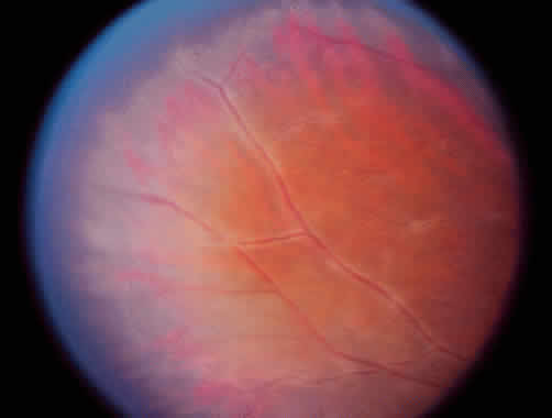
“Uveitis is the medical term we use to describe inflammation of the middle or internal layers of the eye. The symptoms of uveitis vary depending on if the inflammation involves the anterior or front of the eye or the posterior or back of the eye. Common symptoms include, redness of the eye, blurred vision, sensitivity to light or photophobia, floating spots in the visual field, eye pain, floaters, headaches, injected conjunctiva. When we look in the eyes we often see dilated blood vessels, presence of cells in the eye, and precipitates on the surface of the cornea. If the inflammation is severe there may be evidence of a hypopyon or a layer of pus in the eye (see picture below). Inflammation at the back of the eye generally does not have any of these typical symptoms and is more difficult to assess.”

“Uveitis is a common condition and has many causes. In the context of MS it is considered an autoimmune disease. Up to 30% of MSers get uveitis and therefore it is sensible for an ophthalmologist to assess MSers with visual symptoms to exclude uveitis. Another associated disease which is classified as being uveitis is posterior venulitis, when you see inflammation around the veins in the retinae. This inflammation is considered to be the same inflammation we see around veins in other MS lesions. The big difference is the eye or retina does not normally have any myelin. This implies that the inflammation cannot be in response to myelin, but some other antigen. This is another nugget of gold that challenges the dogma that MS is an autoimmune disease targeting myelin.”
 |
| The white sheathing around the blood vessels of the retinae is called retinal vasculitis and is occurs in up to a third of MSers. This is telling us that what ever is driving inflammation in MS may also be present in the layer of the retinae. It is not myelin and the nerve fibres in the retina don’t have myelin. Interesting? |
Before you write in to remind Prof GWhilst there may be no myelin in the eye, autoimmunity to myelin basic protein has been reported to induce uvieitis in the rat. So food for thoughtAdamus G, Sugden B, Arendt A, Hargrave PA.Importance of cryptic myelin basic protein epitopes in the pathogenicity of acute and recurrent anterior uveitis associated with EAE.J Neuroimmunol. 2001;113:212-9.de Vos AF, Dick AD, Klooster J, Broersma L, McMenamin PG, Kijlstra A. Analysis of the cellular infiltrate in the iris during experimental autoimmune encephalomyelitis.Invest Ophthalmol Vis Sci. 2000;41:3001-10.Adamus G, Amundson D, Vainiene M, Ariail K, Machnicki M, Weinberg A, Offner H.Myelin basic protein specific T-helper cells induce experimental anterior uveitis.J Neurosci Res. 1996;44513-8.etc.
Thanks for the citations. I'd heard about no myelin in the retina but not about autoimmunity to myelin inducing uveitis in rats. I'm in the 30% with uveitis and MS and have wondered what the connection between the two is.
DoctorLove's beau thinks that MS is a response to a lens protien that gets expressed in oligos
Does he know of a specific protein that is expressed in both? That would be quite a lead if it were the antigen for MS. Interesting, thanks
Since oligos are dead prior to white blood cell infiltration, the reaction to that lens protein must come from microglia. If this is the case, then why aren't MS lesions diffuse in all white matter? And then, how do grey matter lesions form?
White blood cell infiltration…microglia are white blood cells and they are resident in adult nervous system, They they are in the pre active lesions with not yet dead oligodendrocytes expressing the stress protein come to the research day and it will be explained.
"Since oligos are dead prior to white blood cell infiltration……….."You state this as if it's beyond doubt. It certainly isn't.
Ok MD, let me rephrase: Since oligos are dead before T & B cells that flow in the bloodstream (and are targets of all newer DMTs), cross the BBB and enter the brain.MD2, correct me if i am wrong: All biopsies of early MS lesions show the same thing, dead oligos in the absence of immune cells from the blood. There is no single early MS lesion documented that has T or B cells or monocytes along with living oligos. So, we are forced to accept, based on available evidence, that all MS lesions start the same way.
Not true VV there are numerous studies with fulminant MS showing T and B cells next to oligodendrocytes. Also the toxic mechanism of action may operate over a distance some way away from infiltrating cells due to the release of toxic mediators that can diffuse from the site of infiltration.You base ALL your hypothesis on one observation from John Prineas. it is likely that the case described was certainly not a typical case of MS if in fact it was MS at all.
Let's be specific: is there a biopsy of an early (less than a day old), not recurrent, not chronic, MS lesion where there are significant populations of T or B cells or macrophages AND the nearby oligos are not yet dead but in the process of dying? Since they are numerous, as you say, can you please provide at least one link?
How exactly would you be able to tell that a new lesion would be less than one day old?
This comment has been removed by the author.
a. The way Prineas measured the 17-hour lesion.b. By consecutive MRIs.Can you provide a link that suggests infiltration of blood immune cells prior to oligodendrocyte apoptosis?
Consecutive MRIs to search for a lesion that wasn't there then was is incredibly speculative (and expensive).BTW to describe the famous Prineas lesion as 17 hours old is misleading. It only means that the patient died 17 hours after disease exacerbation after admittance to hospital. The lesion could have been there for some time before this.This paper is worth a look.http://brain.oxfordjournals.org/content/122/12/2279.full.pdf+htmlYou have to remember that just because T or B cells may not be immediately apparent in some lesions, the release of toxic factors mean they can exert an effect over some distance as well as activating resident microglia.
"Since oligos are dead before T & B cells that flow in the bloodstream (and are targets of all newer DMTs), cross the BBB and enter the brain".You seldom see a sniper next to the dead body…. T and B cells besides cytolytic ones work from distances, likewise can you spot the sniper in a crowd of people, without knowing the killing mechanism is a gun. However, I accept that T cells may not be the killing machine, oligos have no MHC class II so CD4 cells are not going to do it and MHC class I is down regulated not favoring CD8 killing, which could happen if they are infected with virus."MD2 correct me if i am wrong…you are wrong". In the biopsies there were 4 types of lesion according to some Pathologists the oligodendrocyte death lesion was one type-a minor type, you argue for a chronological progression that one becomes the next, or could the lesion have a different aetiology. Even last week I saw two lesions from Hans Lassmeann that he believed had apparent different aetiologies.(b) Consecutive MRI..You are having a laugh to suggest that a 1,3 or even 7T scanner can see a single oligodendrocyte dying..it simply does not have the resolution. However, what MRI does show is that the MRI lesion is generally much bigger than the apparent histological lesion, so just because you can't see something next door does not mean it is not there."Review of the clinical course of the 14-year-old female index case described by Barnett and Prineas is more suggestive of pediatric NMO than pediatric MS. The initial presentation of thoracic transverse myelitis (sensory level at T4) coupled with the fulminant disease course, severe attack-related disability, and final lethal brainstem event with respiratory failure preceded by nausea are more in keeping with NMO than pediatric MS. Furthermore, the lesion location in the brainstem at the floor of the fourth ventricle, an AQP4-rich area, also favors the diagnosis of NMO Bruck et al. Annal Neurol 72:385The pre-active lesions akin to the Prineas lesions have oligodendrocytes and microglia as a central component and no T cells apparently near by. Is the oligodendrocyte always dead, the pathologists do not think so, so people think they have a chance to recover.I keep an open mind.
MD2, "You have to remember that just because T or B cells may not be immediately apparent in some lesions, the release of toxic factors mean they can exert an effect over some distance as well as activating resident microglia."If i get it right, you refer to T and B cells that may be present in other lesions nearby. The problem with this explanation is that it can not explain the formation of the first MS lesion. You would have to find a different mechanism to account for it. Moreover, even if there was such a mechanism, then MS lesions would develop around some specific starting points. Instead, they seem to appear quite randomly within the white matter, retaining though their periventricular distribution. Which means two things: a) the mechanism you propose does not have macroscopic support a) It requires one more, initial way for lesions to form.
MD,"In the biopsies there were 4 types of lesion"These are the 4 types of lesions in general irrespective of their age. However, there is only one type of EARLY lesion, the Prineas type. What's more, Prineas is the one who proposes that all 4 types are different time frames of the same process.Bruck gives us an indirect way to measure the Prineas lesion age, by accepting it as the cause of the respiratory failure. Still, by proposing an NMO diagnosis instead of MS does not make things clearer, since there is yet no clear distinction between NMO and MS lesions. They could very well share a common path. As for the fourth ventricle as an AQP4-rich area… cerebellum is also rich in AQP4, but there are no NMO lesions there.
So what is your point?. Is it a vascular response and infection response?.The vein hypothesis is struggling more and more and Prineas thinks it is tosh
At this stage i am not putting forward any vascular explanation. I'm just trying to show you that your proposition is not enough to explain all the available evidence. The most important part is still missing, in my opinion.
I have iven you my proposition.What stresses/kills the oligos is the important thing.My guess is a virushttp://multiple-sclerosis-research.blogspot.co.uk/2011/12/education-espresso-pathology-lesion.htmlAt the research day maybe will have an possible explanation that brings together both views.
What is important is that DMTs that stop new lesion formation to a large extent stop MS as seen with early treatment with alemtuzumab for example. If oligos dying was the primary event then this would continue unabated no matter what the level of stopping new lesion formation by DMTs so you would expect the MS to get worse no matter what. This doesn't happen with the new DMTs which suggests that immune lesion forming cells must have some role in the process of oligo death.
A friend (not a MSer) struggled with uveitis for some years. Then a doctor started treating her with different antibiotics one after another, finally one of them worked, and the uveitis has not come back since then.Sorry for the off-topic comment but I thought this was most interesting.
That is interesting.
the Uveitis Treatment is multifold. Anti inflammatory drugs such as corticosteroids are given in the form of pills injection or topical eye drops as per need. For treatment of posterior uveitis implantation of a device in the eye may be needed which slowly releases the drugs for up to two and a half years. Antibiotics or antiviral can be given if infection is suspected as the underlining cause.