Regrets

Barts-MS rose-tinted-odometer: ★★

Shortly after leaving Queen Square to take up my current position at Barts and The London Juliet Solomon, a good friend, and one of the research managers who had an office opposite me on the 6th floor of the Institute of Neurology sent me a signed copy of the ‘The Book of Regrets’ she had compiled as a present. She has asked celebrities to write essays on something they had regretted in their lives. The book has become a bestseller with all the proceeds of the sales going to support the National Hospital for Neurology and Neurosurgery. This is the kind of thing Juliet does; she has a very big heart. A mensch!

At the time I thought about what I would write if I was asked to contribute a chapter to the next edition of the book. I am still not sure, but from a professional perspective, my biggest regret is not being more proactive in derisking alemtuzumab as a treatment for MS. It has become clear to me that a small proportion of people with MS (pwMS) who are treated with alemtuzumab and HSCT early in the course of their disease are cured of MS when you use a contemporary definition of an MS cure

If the infusion reactions, infections and secondary autoimmunity problems went away who would choose anything but alemtuzumab to treat their MS?

Infusion reactions: Can we reduce the infusion reactions of alemtuzumab? Yes, we can. Pre-treating with steroids starting the night before and moving from the intravenous to the subcutaneous route would make infusion reactions minor. So why hasn’t this been done? Money! MS centres/units make lucre out of infusing patients. I have used the subcutaneous route to avoid a second about of steroid-induced psychosis, to avoid steroid-induced metabolic mayhem in a patient with MS and type 1 diabetes and in a patient who developed avascular necrosis of one hip after his first course of alemtuzumab. It was remarkable; there were no major alemtuzumab infusion reactions despite these three patients being steroid-free. The pharmacodynamic data, i.e. the cell depletion data for the IV and SC routes are identical. The main reasons for us not switching all our patients to subcutaneous alemtuzumab was money, resource and inertia. In the recent past, we used to make money for the unit by giving infusions. Fortunately, with NHS block contracts, this perverse incentive has disappeared. Sanofi-Genzyme also provides contract nurses who come in to give the infusions and monitor the patients. If we converted our entire alemtuzumab administration programme to a subcutaneous route the workload would fall on our overworked nurses. Our nursing lead in our daycare unit reminded me of this. REGRET 1 we didn’t covert to sc alemtuzumab. 

Secondary autoimmunity: What about preventing or reducing the incidence of secondary autoimmunity?

The immune system has many mechanisms in place to prevent autoimmunity. When you learn how the immune system works it is really quite surprising that autoimmunity is so uncommon. What the immunologists tell us is that there must be a series of underlying biological processes that are causing secondary autoimmunity and if we can work out what these are we can intervene and prevent this complication. This is what Joanne Jones and Alasdair Coles tried to do in Cambridge. Their hypothesis was that because the immune system reboots itself from peripheral memory cells it is more likely to result in an aberrant autoimmune response. They tried rebooting the immune system using more naïve cells from the thymus using the hormone palifermin, which stimulates the thymus to produce more naïve T-cells. Sadly it didn’t work, but at least they tried and they should be congratulated for doing this study.  

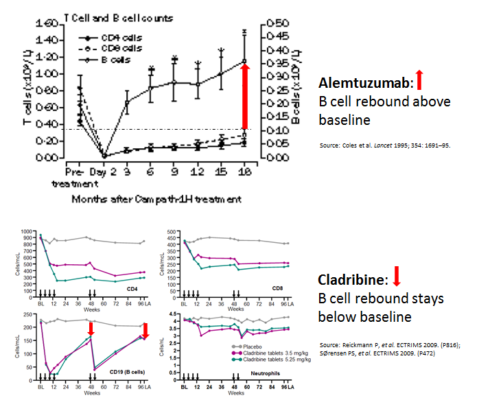
Interestingly, when you compare cladribine, another IRT, with alemtuzumab you can’t help but notice that the B-cell reconstitution profiles are very different. With alemtuzumab, they come back very quickly and overshoot their baseline values. We, and others, have hypothesised that if you change the profile of the B cell reconstitution with a small dose of the B cell depleting antibody rituximab you may be able to prevent this secondary autoimmunity. We are really talking about a very small dose of rituximab, i.e. 10-20mg, just enough to delay B cell reconstitution by 4-6 months. I proposed this concept to Genzyme 8 or 9 years ago, but without data to support the hypothesis they were not keen to support an exploratory study. Why didn’t I push to get this funded from another source? If we had done this study back then we would have had the results by now. Can you imagine how impactful this could be for pwMS if we could prevent secondary autoimmunity post-alemtuzumab? REGRET 2 no doing alemtuzumab-rituximab trial and not setting up an adaptive trial platform for testing multiple strategies to prevent secondary autoimmunity post-alemtuzumab.  

Infections: One success story has been the derisking of alemtuzumab-associated infections with prophylactic antibiotics and antivirals, and the proactive approach to baseline infectious disease screening and vaccination. SUCCESS 1 reducing alemtuzumab-associated infections. 

Anti-drug antibodies: Another success story has been exposing alemtuzumab’s problems with anti-drug antibodies (ADAs) and the development of an assay to screen for these antibodies. Why use a therapy, at great expense, that is not going to work because of neutralizing anti-drug antibodies. SUCCESS 2, anti-drug antibody screening.

A big problem that emerged was how alemtuzumab was licensed and used in the USA. The FDA has essentially licensed alemtuzumab with hand-cuffs, therefore, alemtuzumab was and is used as the DMT of last resort in the US. This led to it being used in an older more advanced cohort of pwMS who had comorbidities. In this group of patients, a new adverse event profile emerged, particularly vascular complications. This led to a safety review and the license of alemtuzumab’s use was changed and it is now only used infrequently, second or third -line and often in people with more advanced MS.  I was personally involved with the original EMA submission; it was a real uphill battle to get alemtuzumab licensed as first-line therapy. Allowing the EMA to change how we use alemtuzumab, i.e. making pwMS have to wait to become eligible for the therapy is a travesty. We, Genzyme and MS community, should have made a more robust argument to the CHMP not to change alemtuzumab’s label. REGRET 3 not allowing alemtuzumab to be used first-line in active MS; it can only be used as a first-line agent in patients with rapidly evolving severe (RES) MS (two disabling attacks in a 12 month period). The problem is that very few patients have RES MS as they tend to be treated now before they have their second disabling relapse.  

Finally, my colleagues. Apart from a small group of MSologists, and we know who we are, most MSologists don’t prescribe alemtuzumab. They find the therapy too difficult and risky to use. I have tried to educate and get more of my colleagues to at least offer alemtuzumab as an alternative to HSCT, but to no avail.  In the UK, we were all geared up to do a head-2-head study of alemtuzumab vs. AHSCT. However, once ocrelizumab was licensed the MS community said they would not be able to recruit for this trial so it has now been converted into alemtuzumab or ocrelizumab vs. AHSCT trial. In reality, this study is going to be a head-2-head of ocrelizumab vs. AHSCT study. Not getting the wider MS community to understand how effective alemtuzumab is REGRET 4. Instead of success, we have a generation of refuseniks

The question we need to ask ourselves is do we really want to throw the baby out with the bathwater? We have two, and possibly three, treatment strategies that may cure a minority of pwMS of having MS. Yes, CURE. However, alemtuzumab and HSCT are on the fringe of MS treatments. Why? 

I suppose you are asking about the third option. It may be cladribine. The results of the ORACLE trial of cladribine in patients with clinically isolated syndromes (CIS) are quite remarkable. We are trying to recall the patients from the ORACLE study a decade or more later to see how many are still in remission and haven’t converted to MS. The problem we have is that cladribine is not even a treatment option for CIS despite this stunning data, hence we may be denying a large number of people with CIS, a relatively safe immune reconstitution therapy or IRT, that may prevent many of them from developing MS. The downside of this is the depressing fact that many MSologists still don’t treat CIS (see my blog post on watchful waiting).

Perumal et al. Subcutaneous administration of alemtuzumab in patients with highly active multiple sclerosis. Mult Scler. 2012 Aug;18(8):1197-9.

Alemtuzumab is an anti-CD52 monoclonal antibody with remarkable efficacy in relapsing multiple sclerosis (MS). In clinical trials and off-label use in MS, alemtuzumab has been administered intravenously (IV). Alemtuzumab is approved for chronic lymphoid leukemia as IV. Oncology guidelines recommend alemtuzumab subcutaneous (SC) over IV. There is no report of alemtuzumab SC in MS. We report two patients with highly active relapsing MS who were treated with SC alemtuzumab, had significant improvement and tolerated SC alemtuzumab well without the typical infusion-associated adverse events. SC alemtuzumab in MS warrants further studies as this may enhance patient convenience and minimize infusion-associated adverse events.

Leist et al.  Effect of oral cladribine on time to conversion to clinically definite multiple sclerosis in patients with a first demyelinating event (ORACLE MS): a phase 3 randomised trial. Lancet Neurol. 2014 Mar;13(3):257-67. 

Background: Patients who develop relapsing-remitting multiple sclerosis (MS) present with a first clinical demyelinating event. In this double-blind, multicentre, randomised, phase 3 study we investigated the effect of oral cladribine on conversion to clinically definite MS in patients with a first clinical demyelinating event, when given at the same doses shown to be effective in relapsing-remitting MS.

Methods: Between Oct 21, 2008, and Oct 11, 2010, we recruited patients aged 18-55 years, inclusive, from 160 hospitals, private clinics, or treatment centres in 34 countries. Eligible patients had a first clinical demyelinating event within 75 days before screening, at least two clinically silent lesions of at least 3 mm on a T2-weighted brain MRI scan, and an Expanded Disability Status Scale score of 5.0 or lower. Patients with a first clinical demyelinating event ≤75 days before screening were randomly assigned (1:1:1) to receive cladribine tablets at cumulative doses of 5.25 mg/kg or 3.5 mg/kg or placebo. Randomisation was done with a central web-based randomisation system and was stratified by geographic region. Masking was maintained using a two-physician model. The primary endpoint of this 96-week study was time to conversion to clinically definite MS according to the Poser criteria. This study is registered with ClinicalTrials.gov, number NCT00725985.

Findings: Of 903 participants assessed for eligibility, 616 patients received cladribine 5.25 mg/kg (n=204), cladribine 3.5 mg/kg (n=206), or placebo (n=206). At trial termination on Oct 25, 2011, cladribine was associated with a risk reduction versus placebo for time to conversion to clinically definite MS (hazard ratio [HR] for 5.25 mg/kg=0.38, 95% CI 0.25-0.58, p<0.0001; HR for 3.5 mg/kg=0.33, 0.21-0.51, p<0.0001). Adverse events were reported in 165 (81%) patients in the cladribine 5.25 mg/kg group, 168 (82%) patients in the cladribine 3.5 mg/kg group, and 162 (79%) patients in the placebo group. We noted no increase in risk of adverse events with active treatment versus placebo apart from lymphopenia, which was a severe event in 10 (5%) patients in the 5.25 mg/kg group and four (2%) patients in the 3.5 mg/kg group.

Interpretation: Both doses of cladribine significantly delayed MS diagnosis compared with placebo. The safety profile of cladribine was similar to that noted in a trial in patients with relapsing-remitting MS. Further research could clarify the potential effects of oral cladribine treatment in the early stages of MS.

Conflicts of Interest

Preventive Neurology

Twitter

LinkedIn

Medium

General Disclaimer: Please note that the opinions expressed here are those of Professor Giovannoni and do not necessarily reflect the positions of the Barts and The London School of Medicine and Dentistry nor Barts Health NHS Trust.

Two, four, six, eight, who do you appreciate?

Barts-MS rose-tinted-odometer: ★

This week I was asked by two patients independently of each other who had delayed access to treatment if this could explain why they now have secondary progressive MS. In parallel, I had a meeting with a friend yesterday, who was the primary driver of the 21-year interferon-beta follow-up survival study (see below), who said that to him this was the most convincing data to support the early treatment paradigm in MS. 

In summary, trial subjects in the pivotal original interferon-beta-1b study who had delayed access to interferon-beta-1b because they were randomised to receive placebo were 50% less likely to be alive at 21 years compared to study subjects with early access to treatment. Was this due to the impact of interferon-beta on MS or some other confounding factor? In the 69 pwMS for whom information on the relationship of death to MS was available 78% were judged to have died from MS-related complications. This tells us that IFN-beta increases your life expectancy by reducing MS-related complications that can cause death in the future. The latter include swallowing problems that are associated with aspiration pneumonia, urinary dysfunction that lead to urinary tract infections and septicaemia, immobility and pressure sores, falls and fractures, to name the most common causes of premature death in pwMS. 

You need to remember that this was a placebo-controlled study and the subjects on placebo were switched to active treatment after 3 years, with some subjects having to wait up to 5 years (trial recruitment was from June 1988 to May 1990; with placebo-treated subjects given free commercial supply as of October 1993). What this study shows is that delaying access to treatment by just 3-5 years has a major impact on long-term outcomes. 

Please note that apart from suicide most pwMS get to EDSS 10 or death as a result of MS by passing through EDSS stages 2, 4, 6 (stick), 8 (wheelchair/bed). So my answer to these patients was yes your delayed access to DMTs in the early 2000s is almost certainly the reason why you now have secondary progressive MS and are disabled. 

Please note this there is now overwhelming evidence from other controlled trials and real-life data to support the interferon-beta 21-year mortality data. In fact, the debate about early treatment and MS outcomes is so widely accepted that it has now has shifted from early access to DMTs to early access to highly effective DMTs, i.e. flipping the pyramid. It is clear that pwMS who are treated with more effective DMTs first-line do so much better than those who are asked to wait (watchful waiting) or are escalated gradually up the DMT ladder (slow escalation). This is why, despite us designing the early treat-2-target NEDA trial, our centre couldn’t participate in the trial comparing maintenance-escalation vs. flipping the pyramid. We simply don’t have equipoise; in other words, we don’t think it is ethical to randomise patients to a treatment arm that is less effective and hence will result in poorer long-term outcomes and probably poorer long-term survival. This is why it is our current clinical practice to offer pwMS who have active disease and are eligible under the NHSE algorithm for treatment access to highly effective treatment first-line. 

We are so convinced about the early treatment that we are doing the ATTACK-MS trial, which will explore if access to the highly effective treatment natalizumab, 2 months earlier than what happens in routine practice, improves outcomes. Yes, we will be randomising patients before they have finished the MS diagnostic pathway to treatment vs. delayed access, i.e. only 2 months later when they have a definitive diagnosis of MS. We anticipate that the ATTACK-MS study will activate the MS community to treat the MS brain as we treat the brain in stroke. Forget about years, every day, week or month for the untreated MS brain is like every second, minute or hour in the non-perfused brain of a person having a stroke.

Yes, in MS time really is brain and by delaying access to DMTs and potentially highly effective DMTs is unacceptable in the modern era of treating MS. The data below not only supports the maxim “Time is Brain” but “Time is Life“, why would anyone wait to treat someone with active MS knowing this?

Figure from Neurology 2012;78(17):1315-22.

Goodin et al. Survival in MS: a randomized cohort study 21 years after the start of the pivotal IFNβ-1b trial. Neurology. 2012 Apr 24;78(17):1315-22.

Objective: To examine the effects of interferon beta (IFNβ)-1b on all-cause mortality over 21 years in the cohort of 372 patients who participated in the pivotal randomized clinical trial (RCT), retaining (in the analysis) the original randomized treatment-assignments.

Methods: For this randomized long-term cohort study, the primary outcome, defined before data collection, was the comparison of all-cause mortality between the IFNβ-1b 250 μg and placebo groups from the time of randomization through the entire 21-year follow-up interval (intention-to-treat, log-rank test for Kaplan-Meier survival curves). All other survival outcomes were secondary.

Results: After a median of 21.1 years from RCT enrollment, 98.4%(366 of 372) of patients were identified, and, of these, 81 deaths were recorded (22.1% [81 of 366]). Patients originally randomly assigned to IFNβ-1b 250 μg showed a significant reduction in all-cause mortality over the 21-year period compared with placebo (p = 0.0173), with a hazard ratio of 0.532 (95% confidence interval 0.314-0.902). The hazard rate of death at long-term follow-up by Kaplan-Meier estimates was reduced by 46.8% among IFNβ-1b 250 μg-treated patients (46.0% among IFNβ-1b 50 μg-treated patients) compared with placebo. Baseline variables did not influence the observed treatment effect.

Conclusions: There was a significant survival advantage in this cohort of patients receiving early IFNβ-1b treatment at either dose compared with placebo. Near-complete ascertainment, together with confirmatory findings from both active treatment groups, strengthens the evidence for an IFNβ-1b benefit on all-cause mortality.

Classification of evidence: This study provides Class III evidence that early treatment with IFNβ-1b is associated with prolonged survival in initially treatment-naive patients with relapsing-remitting multiple sclerosis.

Goodin et al. Cause of death in MS: long-term follow-up of a randomised cohort, 21 years after the start of the pivotal IFNβ-1b study. BMJ Open. 2012 Nov 30;2(6).

Objectives: Compared with controls, multiple sclerosis (MS) patients die, on average, 7–14 years prematurely. Previously, we reported that, 21 years after their participation in the pivotal randomised, controlled trial (RCT) of interferon β-1b, mortality was reduced by 46–47% in the two groups who received active therapy during the RCT. To determine whether the excessive deaths observed in placebo-treated patients was due to MS-related causes, we analysed the causes-of-death (CODs) in these three, randomised, patient cohorts.

Design: Long-term follow-up (LTF) of the pivotal RCT of interferon β-1b.

Setting: Eleven North American MS-centres participated.

Participants: In the original RCT, 372 patients participated, of whom 366 (98.4%) were identified after a median of 21.1 years from RCT enrolment.

Interventions: Using multiple information sources, we attempted to establish COD and its relationship to MS in deceased patients.

Primary outcome: An independent adjudication committee, masked to treatment assignment and using prespecified criteria, determined the likely CODs and their MS relationships.

Results: Among the 366 MS patients included in this LTF study, 81 deaths were recorded. Mean age-at-death was 51.7 (±8.7) years. COD, MS relationship, or both were determined for 88% of deaths (71/81). Patients were assigned to one of nine COD categories: cardiovascular disease/stroke; cancer; pulmonary infections; sepsis; accidents; suicide; death due to MS; other known CODs; and unknown COD. Of the 69 patients for whom information on the relationship of death to MS was available, 78.3% (54/69) were adjudicated to be MS related. Patients randomised to receive placebo during the RCT (compared with patients receiving active treatment) experienced an excessive number of MS-related deaths.

Conclusions: In this long-term, randomised, cohort study, MS patients receiving placebo during the RCT experienced greater all-cause mortality compared to those on active treatment. The excessive mortality in the original placebo group was largely from MS-related causes, especially, MS-related pulmonary infections.

If you are interested you can watch my presentation from last year’s ACTRIMS-ECTRIMS meeting during which I make the case for flipping the pyramid. Please note the ATTACK-MS study paradigm takes this concept of time is brain even further. Do you agree with this approach?

Conflicts of Interest

Preventive Neurology

Twitter

LinkedIn

Medium

General Disclaimer: Please note that the opinions expressed here are those of Professor Giovannoni and do not necessarily reflect the positions of the Barts and The London School of Medicine and Dentistry nor Barts Health NHS Trust.

The Star-MS trial: to be HSCTed or not

Barts-MS rose-tinted-odometer: ★★★

There is little doubt in my mind that HSCT is the most effective DMT we have for treating MS. The latter is based on NEDA (no evident disease activity) rates post-HSCT and brain volume loss data after year one. HSCT appears to put the majority of treated patients into long-term remission and normalises end-organ damage as measured by brain volume loss. In addition, a not insignificant proportion of HSCT-treated patients may be cured depending on how you want to define an MS cure. Despite these data most MSologists however, consider HSCT too risky to refer patients for treatment and hence prefer to go with the more acceptable risk profile of licensed DMTs. This is why HSCT hasn’t taken off as a mainstream treatment for MS and remains a niche treatment. 

What is not known is that HSCT may yet prove to be one of the most cost-effective DMTs we have. Although quite expensive, with most of the costs front-loaded, HSCT does lead to significant cost savings in the long-term (see study below). I wonder if healthcare systems will clock this and take the bold step of underwriting more HSCT treatments for MS with the promise of long term cost savings? The problem we have in medicine is that healthcare budgets typically run on an annual cycle and so costs savings unless made the same year often don’t influence treatment decisions. 

“Why should I spend more money today to only save money in the 5 years time? My responsibility is to this year’s or maybe next year’s budget not the budget in 5 years time.”

The counter-argument to this is should be if I don’t save my brain volume this year or next year, by the time I get to year 5 in my disease course it will be too late. At the moment MS brain and spinal cord damage are irreversible. Yes, time really is brain.

What do you do as a person with MS who has decided to be treated with HSCT, but your neurologist says no? Do you find a neurologist who will say yes? Do you travel abroad and take the private route? Or do you accept your neurologist’s advice and go for the safer, but ultimately more expensive, licensed but less effective DMT? 

The good news is that we in the UK will soon be starting the StartMS trial, which will compare autologous stem cell transplantation (AHSCT) versus alemtuzumab or ocrelizumab in relapsing-remitting MS. This means at least some of you will be offered the opportunity to be randomised to AHSCT or ocrelizumab/alemtuzumab. A major outcome of this study will be a cost-comparison to see how much money it will save the NHS. Exciting or not? Some people are arguing that we don’t need this study as the information is already available. Not sure I agree. Sometimes doing your own research and generating your own data is what is required to change behaviour, i.e. the wide adoption of HSCT as a treatment for MS. 

Orlewska et al. Impact of Immunoablation and Autologous Hematopoietic Stem Cell Transplantation (AHSCT) on Treatment Cost of Multiple Sclerosis: Real-World Nationwide Study. Value Health Reg Issues. 2021 Apr 14;25:104-107.

Objectives: To provide real-world data on the impact of autologous hematopoietic stem cell transplantation (AHSCT) on treatment costs of patients with multiple sclerosis (MS) in Poland.

Methods: Medical data of 105 patients who underwent AHSCT in the years 2011 to 2016 were obtained from the National Health Fund (NHF) database. Treatment costs were calculated from the public payer’s perspective per patient-year for the total available period as well as 12 months before and after AHSCT. The statistical analysis was performed using MATLAB 2016b.

Results: Mean treatment-related costs covered by the NHF per patient-year before and after the transplantation were €4314.9 and €1188.8 , respectively. The average cost of disease-modifying drugs per patient was reduced from €2497.9/year before to €65.3/year after AHSCT.

Conclusions: Although the initial cost of AHSCT is high, the costs involving AHSCT and post-AHSCT treatment could, according to our analysis, pay off in 3.9 years, when compared to the costs of disease-modifying drug therapy in aggressive MS. The study provides evidence that the AHSCT can lead to significant savings in treatment costs of aggressive MS from the public payer’s perspective.

Conflicts of Interest

Preventive Neurology

Twitter

LinkedIn

Medium

Disclaimer: Please note that the opinions expressed here are those of Professor Giovannoni and do not necessarily reflect the position of the Barts and The London School of Medicine and Dentistry nor Barts Health NHS Trust.

How soon does the shredder begin to shred?

Barts-MS rose-tinted-odometer: zero-★s

She was only 26-years and she couldn’t understand why she was falling behind her peers at work. She started working at an ultra-competitive law firm after finishing as one at the top of her peer group at Oxford. She was clearly the best-performing intern in the 2017 intake, which is why she was kept on after her internship. However, things were now going wrong. She was suffering from chronic fatigue, forgetfulness and she simply couldn’t juggle the complex tasks she was being expected to working for more than one client at a time. This job was a high-octane one and you were expected to perform at the level. Her poor performance and increasing list of mistakes had resulted in one performance review already. What should she do?

The back story to this young lawyer is her identical twin sister had been diagnosed with MS at the age of 18, shortly after completing her A-levels. Her sister had decided to delay going to university because of being diagnosed with MS. The odds are this young lawyer had asymptomatic MS and her fatigue and cognition problems are linked to smouldering MS. Do you think she should seek a neurological opinion? She is aware that her lifetime risk of being diagnosed with MS is about 30%.

Do you think she should seek a neurological opinion?

I have made the case that the real MS is not relapses and/or focal MRI activity, but smouldering MS. The real question is when do the pathological processes that drive smouldering MS begin? In this study on asymptomatic MS (radiologically-isolated syndrome or RIS) a third of them already have cognitive impairment and two-thirds had lesions with paramagnetic rims (PRL), i.e. a rim of hot microglia. These so-called PRLs are the precursor to the dreaded SELs (slowly-expanding lesions) that are so unresponsive to our current treatments and responsible for so much damage in MS. 

So what are the implications of this study for MS? 

  1. MS begins long before your first attack.
  2. Smouldering MS, formerly known as progressive MS, also begins long before your first attack.
  3. PRLs and SELs, one of the substrates for smouldering MS, are part of MS pathology from very early in the disease course; possibly the beginning.
  4. Cognitive impairment and end-organ damage begin very early in the course of MS.
  5. We need to change our diagnostic criteria to allow MS to be diagnosed very early on, in this case in the so-called asymptomatic phase of the disease. By using PRLs and the central vein sign (CVS) we are likely to improve the sensitivity and specificity of the diagnostic criteria. So what are we waiting for?

We clearly need a new treatment paradigm to tackle smouldering MS. The current anti-inflammatory monotherapy model of treating MS is unlikely to work. We need combination therapies ASAP. To achieve the latter we are going to have to get Big Pharma and the regulators to innovate quickly and intelligently. 

Oh et al. Cognitive impairment, the central vein sign, and paramagnetic rim lesions in RIS. Mult Scler. 2021 Mar 23:13524585211002097.

Objective: The central vein sign (CVS) and “paramagnetic rim lesions” (PRL) are emerging imaging biomarkers in multiple sclerosis (MS) reflecting perivenular demyelination and chronic, smoldering inflammation. The objective of this study was to assess relationships between cognitive impairment (CI) and the CVS and PRL in radiologically isolated syndrome (RIS).

Methods: Twenty-seven adults with RIS underwent 3.0 T MRI of the brain and cervical spinal cord (SC) and cognitive assessment using the minimal assessment of cognitive function in MS battery. The CVS and PRL were assessed in white-matter lesions (WMLs) on T2*-weighted segmented echo-planar magnitude and phase images. Multivariable linear regression evaluated relationships between CI and MRI measures.

Results: Global CI was present in 9 (33%) participants with processing speed and visual memory most frequently affected. Most participants (93%) had ⩾ 40% CVS + WML (a threshold distinguishing MS from other WM disorders); 63% demonstrated PRL. Linear regression revealed that CVS + WML predicted performance on verbal memory(β =-0.024, p = 0.03) while PRL predicted performance on verbal memory (β = -0.040, p = 0.04) and processing speed (β = -0.039, p = 0.03).

Conclusions: CI is common in RIS and is associated with markers of perivenular demyelination and chronic inflammation in WML, such as CVS + WML and PRL. A prospective follow-up of this cohort will ascertain the importance of CI, CVS, and PRL as risk factors for conversion from RIS to MS.

CoI: multiple

Twitter: @gavinGiovannoni                                              Medium: @gavin_24211

St Elsewhere

Barts-MS rose-tinted-odometer: zero ★’s

St Elsewhere is a euphemism in medicine for a fuck-up done under someone else’s watch, i.e. at St Elsewhere’s hospital. In this week’s NEJM there is a tragic case of a 22-year old Pakistani man who was misdiagnosed as having MS at St Elsewhere, started on natalizumab, which was ineffective, before being switched to interferon beta. By the time he was admitted to UCSF he was in extremis. A relatively standard workup revealed this gentleman had a tumour; a CNS germinoma that had spread to the meninges or coverings of the brain and spinal cord. On reviewing his initial presentation there were so many ‘red flags’ it is hard to understand why he was misdiagnosed. Tragically this poor man now has ‘substantial neurologic disability, for which he received assistance with all activities of daily living’. This case is a tragedy because CNS germinomas when caught early and treated have a reasonable prognosis with a 10-year survival rate of about 70%. 

Minter et al. Stalking the Diagnosis. N Engl J Med. 2021 Apr 1;384(13):1262-1267. 

If only this patient had had a lumbar puncture and CSF analysis done as part of his initial diagnostic work-up the correct diagnosis would have been made. The question arises whether or not this gentleman had medical insurance or not? He was a delivery driver and had only recently immigrated to the USA. To be fair to the neurological team looking after him a diagnostic short-cut may have had to be made because he simply didn’t have the financial resources to pay for the diagnostic tests. I suspect the latter is likely to have happened and maybe the title of this post should have been St Poor.

I want to remind you that if you have been diagnosed with MS you may not have MS. In the study below approximately 1 in 5 people diagnosed with MS don’t have MS. This figure is much higher than previous studies. I have always quoted the Danish post-mortem studies that suggest that about 1 in 20 patients are misdiagnosed. Maybe Danish neurologists are simply better at diagnosing MS compared to their American colleagues?

There is no one test that can be done to diagnose MS. MS is diagnosed by combing a set of clinical and MRI findings, electric or neurophysiological investigations and laboratory tests. If these tests fulfil a set of so-called MS diagnostic criteria the Healthcare professional (HCP) or neurologist makes a diagnosis of MS.

The underlying principle of making a diagnosis of MS is showing dissemination of lesions in space and time and excluding other possible diagnoses that can mimic MS. The diagnostic criteria have evolved over time from being based purely on clinical attacks to those including electrical and spinal fluid tests to the modern era in which we use MRI to help confirm dissemination in time and space.

Dissemination in time means at least two attacks or two MS lesions occurring at least 30 days apart. 

Dissemination in space means lesions occurring in different locations, for example, the optic nerve and spinal cord.

The electrical or neurophysiological tests are called evoked potential (EPs) and test electrical conduction in a particular neuronal pathway. They can be useful to show the effects of lesions in pathways that are not evident on neurological examination or seen on MRI. The EPs can also show slow electrical conduction which is one of the hallmarks of diseases that affect myelin, the insulation of nerves that are responsible for speeding up electrical conduction.

The laboratory tests are typically done to exclude other diseases that can mimic MS. One test that is useful in helping to make the diagnosis of MS is examining the spinal fluid for the presence of oligoclonal immunoglobulin G or IgG bands (OCBs), which are the fingerprint of a specific type of immune activation within the central nervous system (CNS). The OCB fingerprint is relatively specific for the diagnosis of MS in the correct clinical context. Please note OCBs can are found in infections of the nervous system and other autoimmune diseases, therefore, the presence of OCBs are not diagnostic on their own. 

When CSF is sent to the laboratory they also measure the protein, glucose, lactose and do a cell count. An often the spins the cells out of the CSF and examine them to make sure they are abnormal. I suspect if this patient had had a CSF examination it may have been abnormal, which would have led the clinicians to the correct diagnosis, which will have allowed him to be treated differently; importantly, treated early and he may not have become profoundly disabled. 

Why is getting the correct diagnosis of MS so important? Firstly, some of the treatments for MS have life-threatening complications; you don’t want to expose people without MS to these complications. Some diseases that mimic MS can be made worse by MS DMTs. This latter is particularly relevant for NMO or neuromyelitis optic. Patients with NMO misdiagnosed as having MS get worse on many of the MS DMTs. Finally, a diagnosis of MS has many psychological, social, financial and economic implications for people. Just having a diagnosis of MS, even if you turn out to have benign MS in the future, has implications for the person concerned. For example, it may affect your life choices and may impact your ability to get insurance cover to name to obvious examples. I would, therefore, advise you to make sure you have MS and not an MS mimic.

The most common MS mimics:

  1. Cerebrovascular disease
  2. Acute disseminated encephalomyelitis or ADEM
  3. Neuromyelitis optica or NMO
  4. Behcet’s syndrome
  5. Migraine
  6. Sarcoidosis
  7. SLE or systemic lupus erythematosus
  8. Antiphospholipid antibody syndrome
  9. Leukodystrophies

The evolving definition of MS based on diagnostic criteria:

Clinical criteria only:

  1. Schumacher, et al. Problems of Experimental Trials of Therapy in Multiple Sclerosis: Report by the Panel on the Evaluation of Experimental Trials of Therapy in Multiple Sclerosis. Ann N Y Acad Sci 1965;122:552-68.

Clinical, EPs and CSF analysis:

  1. Poser, et al. New diagnostic criteria for multiple sclerosis: guidelines for research protocols. Ann Neurol 1983;13:227-31.

Clinical, EPs, CSF analysis and MRI:

  1. McDonald, et al. Recommended diagnostic criteria for multiple sclerosis: guidelines from the International Panel on the diagnosis of multiple sclerosis. Ann Neurol 2001;50:121-7.
  2. Polman, et al. Diagnostic criteria for multiple sclerosis: 2005 revisions to the “McDonald Criteria”. Ann Neurol 2005;58:840-6.
  3. Polman, et al. Diagnostic criteria for multiple sclerosis: 2010 revisions to the McDonald criteria. Ann Neurol. 2011;69:292-302.
  4. Thompson et al. Diagnosis of multiple sclerosis: 2017 revisions of the McDonald criteria. Lancet Neurol. 2018 Feb;17(2):162-173.

Kaisey et al. Incidence of multiple sclerosis misdiagnosis in referrals to two academic centers. Mult Scler Relat Disord. 2019 May;30:51-56.

BACKGROUND: Multiple Sclerosis (MS) specialists routinely evaluate misdiagnosed patients, or patients incorrectly assigned a diagnosis of MS. Misdiagnosis has significant implications for patient morbidity and healthcare costs, yet its contemporary incidence is unknown. We examined the incidence of MS misdiagnosis in new patients referred to two academic MS referral centers, their most common alternate diagnoses, and factors associated with misdiagnosis.

METHODS: Demographic data, comorbidities, neurological examination findings, radiographic and laboratory results, a determination of 2010 McDonald Criteria fulfillment, and final diagnoses were collected from all new patient evaluations completed at the Cedars-Sinai Medical Center and the University of California, Los Angeles MS clinics over twelve months.

RESULTS: Of the 241 new patients referred with an established diagnosis of MS, 17% at Cedars-Sinai and 19% at UCLA were identified as having been misdiagnosed. The most common alternative diagnoses were migraine (16%), radiologically isolated syndrome (9%), spondylopathy (7%), and neuropathy (7%). Clinical syndromes and radiographic findings atypical for MS were both associated with misdiagnosis. The misdiagnosed group received approximately 110 patient-years of unnecessary MS disease-modifying therapy.

CONCLUSION: MS misdiagnosis is common; in our combined cohort, almost 1 in 5 patients who carried an established diagnosis of MS did not fulfil contemporary McDonald Criteria and had a more likely alternate diagnosis.

CoI: multiple

Twitter: @gavinGiovannoni                                              Medium: @gavin_24211

Why does fingolimod affect antibody responses?

Barts-MS rose-tinted-odometer:  ★

The Twittersphere is abuzz with the preliminary seroconversion rates in Israelis patients with MS on various DMTs in response to the Pfizer-BionTech COVID-19 vaccine (see below). As expected the antibody seroconversion rates in response to anti-CD20 therapies and S1P modulators are blunted and in most cases inhibited. The backstory or biology around anti-CD20 therapy is well-rehearsed; anti-CD20 therapy depletes B-cells and annihilates germinal centres in lymph nodes and the spleen.

The question I have just been asked is why does fingolimod block antibody responses? To answer this we need to go back to the basics of immunology. 

Fingolimod and other S1P modulators work by internalising the S1P receptor making lymphocytes unresponsive to the S1P signalling or chemotaxis gradient in secondary lymph organs such as lymph nodes. In lay language, this causes lymphocytes to park-up in a long-term car park with a wheel lock-on. Even if you wanted to drive your car out of the car park by starting it up you wouldn’t be able to move it without removing the wheel-lock (fingolimod). By blocking lymphocyte mobility helper T-cells can’t migrate to the so-called germinal centres in the lymph nodes and spleen to help B-cells switch from IgM to IgG antibody production and to then help the B-cells to affinity mature their antibodies, i.e. to make good quality antibodies. Normally these affinity matured B-cells would leave the germinal centre to become memory B-cells or plasmablasts. The plasmablasts then mature to become plasma cells and produce high-quality antibodies, which in the case of anti-spike protein protect you from getting COVID-19 in particular severe COVID-19. Fingolimod and other S1P modulators prevent this normal immunology from happening hence the low or absent anti-COVID-19 antibody response after COVID-19 vaccination.

I like to think of the germinal centres as being the immune system’s university; this is where the immune system sends its primed T-cells to help educate B-cells. After a brutal natural selection process in the germinal centres, a few B-cells survive and graduate with a PhD, i.e. a highly specialised degree or class-switched high-affinity IgG antibodies. This then allows the B-cells to become memory B cells and go into semi-retirement or to set-up their own production company as plasma cells and to produce high-quality antibodies. Anti-CD20 therapies work by blowing up the B-cell university and S1P modulators stop the teachers (T-cells) from educating their students (B-cells). Having no university or no teacher-student interactions have the same effect and result in no educated B-cells and hence no IgG antibody responses.

Please note the above information does not change my personal advice regarding vaccination, whether you are on an anti-CD20 therapy, fingolimod or another S1P modulator (siponimod, ozanimod, ponesimod) #GetVaccinated ASAP; some immunity is better than no immunity. Please note having no anti-SARS-CoV-2 antibodies doesn’t necessarily mean you have no immunity. These antibody studies don’t tell us anything about T-cell responses, which are likely to be as important as antibodies in providing protecttive immunity against SARS-CoV-2.

Han et al. FTY720 suppresses humoral immunity by inhibiting germinal center reaction. Blood. 2004 Dec 15;104(13):4129-33. doi: 10.1182/blood-2004-06-2075. Epub 2004 Aug 19.

FTY720 is a novel immunosuppressant that is highly effective in inhibiting rejection of allografts and autoimmunity in various animal models. It has been shown that the sphingosine 1 phosphate (S1P) receptors are the direct molecular targets of FTY720. However, the mechanisms responsible for inhibiting specific immune responses by FTY720 are not well resolved. In particular, there is no available information on whether or how this compound affects humoral immunity. We have investigated the effect of FTY720 treatment on B-cell response during the immune response to a well-defined T-dependent antigen. Our data demonstrated that germinal center reaction was significantly reduced in peripheral lymphoid tissues of mice treated with FTY720. In addition, FTY720 treatment inhibited the production of high-affinity, class-switched antibodies, but not the production of low-affinity, immunoglobulin M (IgM) antibody. Consistently, FTY720 did not have a significant effect on antibody response to a T-independent antigen. Our results may have important implications in application of FTY720 in immune regulation.

Also see the post by MD from a few days ago. Fingolimod also stops B cells moving within the follciles and and stops them contacting areas where they the B cells are likely to be stimulated.

CoI: multiple

Twitter: @gavinGiovannoni                                              Medium: @gavin_24211

Ocrelizumab: DODO vs. ADIOS, who will win?


Barts-MS rose-tinted-odometer: ★★

In response to a question over the weekend about what has happened to the DODO and ADIOS studies. Both are alive and kicking. The more insightful question would be ‘how can I support both the DODO (double-dose ocrelizumab study) and the ADIOS (adaptive dosing ocrelizumab study) studies?’.

Surely, the DODO and ADIOS studies are incompatible with each other scientifically? How can I, on the one hand, support a higher dose of ocrelizumab and on the other hand suggest reducing the dose in the longterm. The hypothesis is all about timing and how you use anti–CD20 therapies.

You need higher doses of anti-CD20 therapy initially as an induction strategy to purge the various B-cell compartments of memory B-cells, which house latent EBV and the highly autoreactive population of B-cells that drive and maintain the MS-state. This population of cells may reside in the deep tissues and/or the central nervous system, which is why we are also testing CNS penetrant anti-B-cell strategies, simultaneously. Time is short so we need to run trials in parallel.

However, once you have purged these compartments say after 2 years of treatment you don’t need to maintain such high-doses of anti-CD20 therapies that are then suppressing normal B-cell biology and immune responses, which result in longterm complications. This is why we want to use ocrelizumab as an immune reconstitution therapy, i.e. high-dose upfront followed by no treatment and wait to see if MS remains in remission or disease-activity returns requiring additional courses. The latter is one of the arms of our proposed ADIOS study. 

In reality, if we could convince a national funding agency, a pharma company or a wealthy philanthropist I would use anti-CD20 therapy as part of an induction-maintenance protocol. After two years of induction therapy with high-dose ocrelizumab, I would test different maintenance strategies in parallel. My agents of choice would be teriflunomide, leflunomide, IMU-838 (vidofludimus) or ASLAN003 (selective second-generation DHODH inhibitors), HAART (highly active antiretrovirals), famciclovir or another anti-EBV viral agent. The hypothesis is to allow B-cell reconstitution after anti-CD20 therapy in the presence of an anti-viral agent to prevent EBV reactivation and reinfection of new memory B cells. By doing this you will also be derisking the long-term immunosuppression associated with anti-CD20 therapies and prevent the development of hypogammaglobulinaemia. In addition, you will be allowing patients to respond to vaccines.

The problem with this trial proposal is the outcome measure; the power calculations are not trivial and the study would have to be very long. I also have reservations about whether or not the regulators will accept the induction maintenance strategy. Maybe we can sell it to them on safety, i.e. to prevent the development of hypogammaglobulinaemia and infections rather than on efficacy? If we go this route then there is only one agent we can use and that is teriflunomide, which is licensed to treat MS. As teriflunomide is coming off patent there is a chance the  NHS may be interesting in funding such a trial; i.e. it would save them money. This is something I am exploring as a proof-of-concept trial.  

The good news is that Roche-Genentech is testing the principles of the DODO study and announced at MSVirtual2020 two high-dose ocrelizumab trials (see below). These trials up the stakes in the anti-CD20 wars and I am confident that we need higher doses upfront to purge deep tissue and possibly CNS pools of B-cells. Please note that you don’t need higher doses of anti-CD20 therapy to suppress relapses and focal MRI activity you can do that with current or lower doses. I am confident both these studies will show that higher-dose ocrelizumab is superior to standard dose ocrelizumab on disability progression or smouldering MS, but not on focal inflammatory events. In relation to the latter, we have hit the ceiling already.

You need higher doses up-front to target the drivers of smouldering MS; i.e. disease progression independent of relapses, accelerated brain volume loss, slowly expanding lesions (SELs) and the subpial cortical lesions.  If these higher-dose studies are positive it will put clear daylight between ocrelizumab and the other anti-CD20 therapies and it would mean the ofatumumab and rituximab are currently being underdosed, at least initially in the first two years. But don’t we have a hint of this already? Ofatumumab was not better than teriflunomide at slowing down brain volume loss in year two of the ASCLEPIOS I and II clinical trials (NCT02792218 and NCT02792231) despite being superior to teriflunomide on relapses and MRI activity. The latter is more proof that focal inflammatory disease (relapses and MRI activity) is not MS but in response to what is causing the disease. The real MS is what causes smouldering pathology and end-organ damage.

DODO vs. ADIOS vs. iTeri: which one would I prioritise? Almost certainly iTeri; the iTeri trial makes the most sense in terms of our current understanding of the pathogenesis of MS, mode of action of anti-CD20 therapies and the long-term risks of chronic B-cell depletion.

Figure from the NEJM.

Hauser et al. Ofatumumab versus Teriflunomide in Multiple Sclerosis. N Engl J Med. 2020 Aug 6;383(6):546-557. 

Background: Ofatumumab, a subcutaneous anti-CD20 monoclonal antibody, selectively depletes B cells. Teriflunomide, an oral inhibitor of pyrimidine synthesis, reduces T-cell and B-cell activation. The relative effects of these two drugs in patients with multiple sclerosis are not known.

Methods: In two double-blind, double-dummy, phase 3 trials, we randomly assigned patients with relapsing multiple sclerosis to receive subcutaneous ofatumumab (20 mg every 4 weeks after 20-mg loading doses at days 1, 7, and 14) or oral teriflunomide (14 mg daily) for up to 30 months. The primary end point was the annualized relapse rate. Secondary end points included disability worsening confirmed at 3 months or 6 months, disability improvement confirmed at 6 months, the number of gadolinium-enhancing lesions per T1-weighted magnetic resonance imaging (MRI) scan, the annualized rate of new or enlarging lesions on T2-weighted MRI, serum neurofilament light chain levels at month 3, and change in brain volume.

Results: Overall, 946 patients were assigned to receive ofatumumab and 936 to receive teriflunomide; the median follow-up was 1.6 years. The annualized relapse rates in the ofatumumab and teriflunomide groups were 0.11 and 0.22, respectively, in trial 1 (difference, -0.11; 95% confidence interval [CI], -0.16 to -0.06; P<0.001) and 0.10 and 0.25 in trial 2 (difference, -0.15; 95% CI, -0.20 to -0.09; P<0.001). In the pooled trials, the percentage of patients with disability worsening confirmed at 3 months was 10.9% with ofatumumab and 15.0% with teriflunomide (hazard ratio, 0.66; P = 0.002); the percentage with disability worsening confirmed at 6 months was 8.1% and 12.0%, respectively (hazard ratio, 0.68; P = 0.01); and the percentage with disability improvement confirmed at 6 months was 11.0% and 8.1% (hazard ratio, 1.35; P = 0.09). The number of gadolinium-enhancing lesions per T1-weighted MRI scan, the annualized rate of lesions on T2-weighted MRI, and serum neurofilament light chain levels, but not the change in brain volume, were in the same direction as the primary end point. Injection-related reactions occurred in 20.2% in the ofatumumab group and in 15.0% in the teriflunomide group (placebo injections). Serious infections occurred in 2.5% and 1.8% of the patients in the respective groups.

Conclusions: Among patients with multiple sclerosis, ofatumumab was associated with lower annualized relapse rates than teriflunomide. (Funded by Novartis; ASCLEPIOS I and II ClinicalTrials.gov numbers, NCT02792218 and NCT02792231.).

CoI: multiple

Twitter: @gavinGiovannoni                                              Medium: @gavin_24211

Remyelination: why have we failed?

Barts-MS rose-tinted-odometer: ★

I sit on several steering committees and advisory boards for projects that are exploring remyelination or recovery of function as a treatment strategy in MS. Doing this allows you to think or groupthink, which has prompted me to consider whether remyelination in MS is necessary or not. I have been scratching my head about why in 2020 three remyelination strategies failed.

  1. High-dose biotin (MedDay) – Effect of MD1003 in Progressive Multiple Sclerosis (SPI2) (ClinicalTrials.gov Identifier: NCT02936037)

    Primary outcome: Proportion of patients Improved on either the EDSS or 25TW
  2. Bexarotene (Cambridge University) – A trial to determine bexarotene’s safety and tolerability and its ability to promote brain repair in patients with multiple sclerosis (EudraCT number 2014-003145-99)

    Secondary or remyelination outcome: Change in mean lesional MTR between month 0 and month 6 for lesions selected for each patient.
  3. Opincinumab (Biogen) – Efficacy and Safety of BIIB033 (Opicinumab) as an Add-on Therapy to Disease-Modifying Therapies (DMTs) in Relapsing Multiple Sclerosis (MS) (AFFINITY) (ClinicalTrials.gov Identifier: NCT03222973)

    Primary outcome: Overall Response Score, a multicomponent score based on 4 components: EDSS, T25FW, 9HPT in the dominant hand and non-dominant hand. 

Have we the MS community made a fundamental mistake in assuming that remyelination is an ongoing persistent problem that needs fixing in MS? Have we got the biology wrong? Is biotin-dependent mitochondrial dysfunction and biotin-dependent remyelination pathways a problem in MS? Is the activation of retinoid X receptors (RXRs) to promote remyelination the correct target in MS? Is the inhibition of an inhibitor of remyelination with opicinumab (anti-LINGO-1) sufficient to promote remyelination across the central nervous system? At least with anti-LINGO-1 or opicinumab, there was proof of biology, i.e. the drug did improve conduction speed in demyelinated optic nerves. 

Were the trial designs correct? For high-dose biotin, the population of pwMS was quite old with a lot of disability. Maybe the population was too old and hence ageing mechanism prevented any recovery of function from being detected. Maybe there was too much axonal or nerve loss and hence remyelination was doomed to fail.

Maybe focusing on the individual MS lesion with bexarotene using MRI techniques is too fickle? It is clear that in the bexarotene study there were too few new, presumably actively demyelinating, lesions to generate a signal. 

The overall response score in the opicinumab trial may not be good enough to capture the recovery of function due to remyelination. The idea of using a composite score to detect recovery of function makes clinical and biological sense. However, using the limbs as the main read-out may not be sensitive enough. What about vision, cognition, balance, bowels, bladder, fatigue, etc. Do we need a better outcome measure? Do we have to go back to the drawing board with our clinical outcome measures?

Or is the answer staring us in the face? When you treat MS early with the big guns, i.e. alemtuzumab or HSCT, it is quite remarkable how many pwMS have spontaneous recovery of function. Are alemtuzumab and HSCT doing something fundamental to the pathogenesis of MS that then allows for spontaneous recovery of function? Are we curing a proportion of people with MS with these treatments?

One of the latest theories that is being tested in MS is that because of premature ageing and senescence the oligodendrocyte precursors and oligodendrocytes (myelin-producing cells) can’t function properly. The solution is to reprogramme them, i.e. dial back the ageing clock by using diet (caloric restriction, intermittent fasting or ketosis) and/or medication (e.g. metformin or fumarates) before hitting them with drugs to stimulate myelin formation. The latter is backed by animal data, but will it work in MS? Possibly; however, this is based on the assumption that the tools we have for measuring remyelination in pwMS actually work.

The danger with all these strategies is tossing the baby out with the bathwater, i.e. once a trial is negative with a compound it is very rare that the community goes back to the compound or biology. So we need to be confident that our methods are sound and reliable. I am not sure we can be that confident after the three failures above. 

Another aspect that is lacking in all our trials is neurorehabilitation, i.e creating the biological stimulus to promote recovery of function. Nobody would perform a spinal cord injury recovery of function trial without active rehabilitation on top of the treatment being tested. The latter is backed up by sound science and compelling animal studies. Why do we in the MS community expect spontaneous recovery of function without rehab? If you don’t use it you lose it! To encourage the recovery of function you need to stress the pathway. I thought this concept would be a no-brainer, but I have yet to convince one Pharma company to add-on a rehab programme to their recovery of function trials.

Maybe instead of trying to promote remyelination and recovery of function in pwMS, our aim should be to treat MS effectively early on, i.e. to prevent the need for remyelination therapies in the first place? The question is how do we get the wider MS community to promote the use of alemtuzumab and HSCT, or similar agents, as the default first-line therapy for MS? Some of us have tried, including me, but have failed miserably with this seemingly easy task. By the time most pwMS get to alemtuzumab or HSCT, they have so much damage that they need restoration therapies. 

As we think we now know the potential cause of MS trying to put out a fire without correcting the upstream pathology is folly. Therefore maybe we should only be trying remyelination and neurorestorative therapies in people who have stable fixed deficits, with no ongoing evidence of any inflammatory disease activity, post alemtuzumab or HSCT? The irony of letting pwMS become disabled before being treated with alemtuzumab or HSCT is inadvertently creating a trial-ready population of subjects for remyelination therapy trials.

I encourage thoughts or counter arguments on the above. Let’s have an open discussion and debate on the points I have raised in this post. I think we need to answer some fundamental questions about remyelination and neurorestorative therapies before we spend more money on futile trials and throw the baby out with the bathwater. 

CoI: multiple

Twitter: @gavinGiovannoni                                              Medium: @gavin_24211

Eight Swallows

Barts-MS rose-tinted-odometer: ★★★★★

How many swallows make a summer? Is eight enough? 

If MS is caused by EBV you would expect there to be clusters of MS potentially linked to a specific subtype of the virus. The best-studied MS cluster is the one from Fjelsø, a small village of 74 families in rural Denmark, where eight people closely linked to each other all developed MS within 13 years of each other. All the subjects had attended the same school. Interestingly all of the people who developed MS had attended the scouts together. 

Danish investigators then typed the variant of EBV these subjects had been infected with and to their surprise, they all had the same subtype of EBV, which importantly was different from controls that were selected from schoolmates and family members. This raises the question of being a scout in Denmark resulted in the transmission of EBV between these subjects. None of these eight subjects reported having had infectious mononucleosis.

What are the chances of getting an eight-person cluster of MS from a group of scouts in a tiny rural village in Denmark? Then on top of this what are the chances of all eight of these people with MS having the same EBV subtype when their family members and schoolmates did not? I suspect the chances are very low.

I don’t think eight swallows are enough to make a summer, but you can’t ignore this cluster when all the other epidemiological evidence points to EBV being causally linked to MS.

The two linked studies below are just a small piece of a large jigsaw puzzle that is gradually being built that I predict will eventually prove EBV is the cause of MS. In the centre of this large jigsaw puzzle are the bespoke pieces for the EBV antiviral and vaccine studies. 

Haahr et al. Cluster of multiple sclerosis patients from Danish community. Lancet. 1997 Mar 29;349(9056):923.

Cluster: We report a cluster of MS in which eight people with verified MS originated from a small Danish community called Fjelsø. All eight had lived within a 2.75 km2 area (2.5 km×1.1 km), where 74 single-family houses, including some farms, were located. The community had a stable population with few migrations into and out of the area. During a 13-year period, all the patients had for 7 years attended the same elementary school with 70-80 pupils. The school had 145 pupils during this period. All those who developed MS had been scouts together, with the older ones being scoutmasters for the younger ones and some of the older ones had also looked after the younger ones. Two cases were siblings and two were aunt and nephew, but MS had not been observed in any of the ancestors of the eight cases or among the school teachers. All cases of MS developed, at various ages and with variable courses, after the eight had left Fjelsø. None of the eight could recall symptoms of infectious mononucleosis.

Munch et al. A single subtype of Epstein-Barr virus in members of multiple sclerosis clusters. Acta Neurol Scand. 1998 Dec;98(6):395-9.

Objectives: Epidemiological studies strongly indicate an infectious involvement in multiple sclerosis (MS). Epstein-Barr virus (EBV), to which all multiple sclerosis patients are seropositive, is also interesting from an epidemiological point of view. We have reported a cluster of MS patients with 8 members from a small Danish community called Fjelsø. To further evaluate the role of EBV in MS we have investigated the distribution of EBV subtypes in cluster members and in control cohorts.

Materials and methods: Blood mononuclear cells were isolated from cluster members, unrelated MS patients, healthy controls, including healthy schoolmates to the Fjelsø cluster patients and finally from persons with autoimmune diseases in order to investigate the number of 39 bp repeats in the EBNA 6-coding region in the EBV seropositive individuals.

Results: We observed a preponderance of the subtype with 3 39 bp repeats in the EBNA 6-coding region both in the MS patients and the healthy controls. In the Fjelsø cluster, all 8 cluster members were harbouring this subtype, which is significantly different from the finding in healthy controls (n = 16), which include 8 schoolmates to the cluster members and 8 randomly selected healthy persons (Fischer’s exact test P = 0.0047), and also compared to all non-clustered individuals studied (P = 0.017).

Conclusion: Infection with the same subtype of EBV links together the 8 persons from the Fjelsø cluster who later developed MS. This finding adds to the possibility that the development of MS is linked to infection with EBV.

CoI: multiple

Twitter: @gavinGiovannoni                                              Medium: @gavin_24211

Swiss-cheese

Barts-MS rose-tinted-odometer: zero-stars

What do you say when I colleague chastises you for stating the fact that MS is potentially a ‘preventable dementia’? This particular colleague was clear that there is no need to draw any parallels between MS and other neurodegenerative diseases because of the negative connations that the term dementia has. I had to remind him that MS ticks all the boxes for being classified as a dementia; i.e. (1) MS is an acquired and not a congenital disorder; (2) MS is a chronic progressive disease; (3) MS impacts on multiple cognitive domains and (4) MS impacts on social and occupational functioning. Have I missed something?

I also had to remind this colleague that almost every neurology or psychiatry textbook included MS on its list of causes of dementia. I am not prepared to peddle alternative facts because pwMS may find it distressing to find out that if MS is left to its own devices it will shred their brains and cause dementia. Please note the rose-tinted-odometer is set to zero for this post. 

The small study below reiterates what we already know that both relapsing and progressive patients have cognitive problems that correlate with physical disability. This study also confirms that T1 hypodensities or blackholes on MRI, particular in the thalamus (a deep grey matter structure in the brain) predicts cognition problems. T1 hypodense MS lesions or black holes, or at least a proportion of them, have been shown to be very destructive and include lesions with so-called phase-rims (iron around them) and a subset we call SELs (slowly expanding lesions). Some neuroradiologists often describe an MS brain with a high volume of black holes as being similar to Swiss cheese in reference to Emmental cheese. 

Now for the good news is that these studies below are on patients with significant end-organ damage and if we can diagnose and treat MS effectively early on we can prevent or at least delay the end-organ damage and the progressive loss of cognition. This is why we have spent years promoting the concepts of ‘Time-is-Brain’, ‘Treat-2-Target of NEDA’, ‘Rapid escalation’, ‘Flipping-the-Pyramid’, ‘Brain Health’, ‘Beyond-NEDA preventing end-organ damage’, ‘Holistic Management’, ‘Marginal Gains’, etc. Buried in all of these concepts is the use of effective DMTs to prevent end-organ damage and to prevent dementia. 

I am very pleased that my pwMS in Australia have taken this one step further and launched their own awareness campaign, albeit sponsored by Biogen, to raise awareness about early effective treatment (www.msmotion.com.au). The campaign is been run by a group of MS social media influencers. I met them all virtually last year and spoke to them about the concepts that underpin our  ‘Brain Health: Time Matters’ policy document. It would be great if pwMS across the world could do a similar thing. 

Do you agree with my colleague above that we should try and protect pwMS from the harsh realities of MS and what can happen to their brains if we don’t manage their MS appropriately? Or should we peddle false facts and a rose-tinted view of the world?

de Paula Gois et al. Associations between cognitive and clinical disability across MS subtypes: The role of the underlying brain damage. Mult Scler Relat Disord. 2020 Dec 19;48:102701. 

Background: Cognitive impairment (CI) is present in all stages and subtypes of multiple sclerosis (MS). However, the majority of studies examined relapsing-remitting (RRMS) patients, and did not address cognitive phenotyping. Is still not clear whether patients with progressive MS (PMS) have a distinct pattern of CI compared to RRMS. In addition, there is conflicting data regarding the correlation between clinical and cognitive disability.

Objective: To investigate the differences of CI between PMS and RRMS patients, evaluating cognitive phenotypes. We also aimed to analyze the association between physical and cognitive disability with MRI measures of grey-matter atrophy and lesion burden.

Methods: Thirty patients with PMS and twenty-four with RRMS underwent neurological, neuropsychological (BRB-N, Boston Naming, and Tower of London), and MRI assessments (3T). Brain volume evaluations were performed using FreeSurfer. Principal Components Analysis on neuropsychological yielded six principal cognitive domains. Cognitive deficits were classified according to three categories: no CI, impairment in isolated cognitive domain, or impairment in combined domains.

Results: In the overall sample, the most frequently impaired cognitive domains were information processing speed (IPS) and visual memory. PMS patients had a higher prevalence of verbal memory and verbal fluency deficits, and more frequent impairment in combined cognitive domains compared to RRMS individuals. After multivariable regression analysis with clinical variables, EDSS was associated with most cognitive domains. Nevertheless, after including T1-lesion volume in the model, it was the most consistent predictor of cognitive performance. To further analyze the interaction between EDSS and T1-lesions, we performed GLM analysis with EDSS and T1-hypointense lesion volume as covariates, and T1-lesion volume adjusted better the model for verbal memory (p = 0.013), IPS (p = 0.021) and total number of impaired cognitive domains (p = 0.021).

Conclusions: RRMS and PMS patients tend to have a similar neuropsychological profile in general, but the extent of CI was greater in PMS patients. Worse cognitive performance was associated with increased physical disability, but this correlation was no longer significant after controlling for T1-lesion volume, suggesting that the underlying MS pathology might be involved in this relationship. Thalamic and T1-lesion volumes were the most consistent MRI predictors associated with cognitive disability.

CoI: multiple

Twitter: @gavinGiovannoni                                         Medium: @gavin_24211

Exit mobile version
%%footer%%