Do you want to be treated with low-dose anti-CD20 therapy?

Barts-MS rose-tinted-odometer: ★★★★★ (rose-red; a climbing rose with thorns)

I have moved my treatment goal beyond NEIDA (no evident inflammatory disease activity) for my patients with MS. The new focus is on preventing end-organ damage. To achieve this we need to take off the blinkers that the Pharma industry has blinded us with. Our treatment target has to be smouldering MS, i.e. stopping disability progression, normalising brain volume loss, flattening neurofilament levels, stop slowly expanding lesions from getting bigger, clearing the CSF of oligoclonal bands and if possible promoting repair and recovery of the nervous system. 

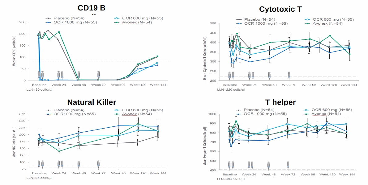
What good is to be free of relapses and focal MRI activity if you are getting worse? This is why the concept of using low dose anti-CD20 therapy is so flawed. It is clear that study subjects exposed to lower doses of ocrelizumab in the phase 3 trials did as well as those exposed to higher doses in relation to relapses and MRI activity, but not in relation to worsening disability (see slideshow below). 

From this post-hoc analysis, it is clear that you need higher, and not lower, doses of anti-CD20 therapy at least initially as an induction strategy to purge the various B-cell compartments. We hypothesise these compartments house memory B-cells, which may be an important sanctuary for latent EBV and/or the highly autoreactive population of B-cells that drive and maintain the MS-state. This population of cells may reside in the deep tissues and/or the central nervous system. This is why we and others are testing CNS penetrant anti-B-cell strategies (ixazomib, cladribine, BTK inhibitors, etc.), i.e. we are going beyond the peripheral B-cell target. 

However, I have hypothesized that once you have purged these compartments, say after 2 years of treatment you may not need to maintain such high doses of anti-CD20 therapy that will then suppress normal B-cell biology and immune responses, which result in long term complications. This is why I have proposed using ocrelizumab as an immune reconstitution therapy, i.e. high-dose upfront followed by no treatment and wait to see if MS remains in remission or disease-activity returns requiring additional courses. The latter is what we are proposing to do in the ADIOS study. 

Even better would be two years of induction therapy with high-dose ocrelizumab followed by a maintenance therapy such as teriflunomide, leflunomide, IMU-838 (vidofludimus) or ASLAN003 (selective second-generation DHODH inhibitors), HAART (highly active antiretrovirals), famciclovir or another anti-EBV viral agent. 

The hypothesis is to allow B-cell reconstitution after anti-CD20 therapy in the presence of an antiviral agent to prevent EBV reactivation and reinfection of new memory B cells. By doing this you will also be derisking the long-term immunosuppression associated with anti-CD20 therapies and prevent the development of hypogammaglobulinemia. This strategy will also allow patients to respond to vaccines.

However, if you want lower dose anti-CD20 therapy you will be able to start Ofatumumab very soon. Please remember ofatumumab was vastly superior to teriflunomide in suppressing relapses and MRI activity (Pharma’s blinkers) but was not superior to  teriflunomide at slowing down brain volume loss in year two of the ASCLEPIOS I and II clinical trials (NCT02792218 and NCT02792231). Why? 

The following is the fundamental question you should ask yourself.

So what would you choose your MS to be treated with; (1) low-dose anti-CD20, (2) high-dose anti-CD20, (3) high-dose anti-CD20 therapy followed by a maintenance treatment or (4) an immune-reconstitution therapy (cladribine, alemtuzumab or AHSCT)?

Sadly we can’t offer all of these choices to all of our patients with MS in the current NHS treatment landscape. 

Figure from the NEJM.

Hauser et al. Ofatumumab versus Teriflunomide in Multiple Sclerosis. N Engl J Med. 2020 Aug 6;383(6):546-557. 

Background: Ofatumumab, a subcutaneous anti-CD20 monoclonal antibody, selectively depletes B cells. Teriflunomide, an oral inhibitor of pyrimidine synthesis, reduces T-cell and B-cell activation. The relative effects of these two drugs in patients with multiple sclerosis are not known.

Methods: In two double-blind, double-dummy, phase 3 trials, we randomly assigned patients with relapsing multiple sclerosis to receive subcutaneous ofatumumab (20 mg every 4 weeks after 20-mg loading doses at days 1, 7, and 14) or oral teriflunomide (14 mg daily) for up to 30 months. The primary end point was the annualized relapse rate. Secondary end points included disability worsening confirmed at 3 months or 6 months, disability improvement confirmed at 6 months, the number of gadolinium-enhancing lesions per T1-weighted magnetic resonance imaging (MRI) scan, the annualized rate of new or enlarging lesions on T2-weighted MRI, serum neurofilament light chain levels at month 3, and change in brain volume.

Results: Overall, 946 patients were assigned to receive ofatumumab and 936 to receive teriflunomide; the median follow-up was 1.6 years. The annualized relapse rates in the ofatumumab and teriflunomide groups were 0.11 and 0.22, respectively, in trial 1 (difference, -0.11; 95% confidence interval [CI], -0.16 to -0.06; P<0.001) and 0.10 and 0.25 in trial 2 (difference, -0.15; 95% CI, -0.20 to -0.09; P<0.001). In the pooled trials, the percentage of patients with disability worsening confirmed at 3 months was 10.9% with ofatumumab and 15.0% with teriflunomide (hazard ratio, 0.66; P = 0.002); the percentage with disability worsening confirmed at 6 months was 8.1% and 12.0%, respectively (hazard ratio, 0.68; P = 0.01); and the percentage with disability improvement confirmed at 6 months was 11.0% and 8.1% (hazard ratio, 1.35; P = 0.09). The number of gadolinium-enhancing lesions per T1-weighted MRI scan, the annualized rate of lesions on T2-weighted MRI, and serum neurofilament light chain levels, but not the change in brain volume, were in the same direction as the primary end point. Injection-related reactions occurred in 20.2% in the ofatumumab group and in 15.0% in the teriflunomide group (placebo injections). Serious infections occurred in 2.5% and 1.8% of the patients in the respective groups.

Conclusions: Among patients with multiple sclerosis, ofatumumab was associated with lower annualized relapse rates than teriflunomide. (Funded by Novartis; ASCLEPIOS I and II ClinicalTrials.gov numbers, NCT02792218 and NCT02792231.).

Conflicts of Interest

Preventive Neurology

Twitter

LinkedIn

Medium

General Disclaimer: Please note that the opinions expressed here are those of Professor Giovannoni and do not necessarily reflect the positions of the Barts and The London School of Medicine and Dentistry nor Barts Health NHS Trust.

Fingolimod vs. Siponimod

Barts-MS rose-tinted-odometer: ★★ (mid-week sepia = #704214)

In my post on rebound disease activity in a person with secondary progressive MS switching from fingolimod to siponimod, someone asked whether there is any logic in switching DMTs within the class of S1P modulators. Two or three years ago I would have said no, but now I would say yes. There are well defined and clear differences between the two compounds that may explain their different effects as DMTs in people with more advanced or progressive MS (see figure and table below). 

The fact that fingolimod works on a broader spectrum of S1P receptors may explain why it has a greater effect on peripheral immune function, i.e. its action on S1P4 may explain why it disrupts antibody responses to new vaccines. S1P4 plays an important role in the functioning of germinal centres (GCs) in lymph nodes and other secondary lymphoid organs, i.e. so-called follicular T-helper cells use S1P4 for migration signals. If these cells can’t enter the GCs they can’t help B-cells make good antibody responses. I, therefore, predict that vaccine responses in response to the COVID-19 vaccines will be better preserved with siponimod, ozanimod and ponesimod because this new generation of S1P modulators has less or no activity on S1P4 receptors.  

Fingolimod needs to be phosphorylated to become active, in comparison siponimod is active already. This may explain why siponimod has greater activity on the S1P5 receptor within the central nervous system (CNS) and explains its greater apparent effects on cells within the central nervous system (CNS). It is clear that when you look at the results of the fingolimod in the PPMS trial there was very little evidence that fingolimod was having any effect on the end-organ, i.e. there was no impact on brain volume loss and no difference across any of the clinical endpoints in the PPMS trial. In comparison, siponimod has a clear CNS signal compared to placebo in subjects with SPMS. Compared to placebo, patients on siponimod have less whole brain, grey matter and thalamic volume loss, preservation of brain tissue integrity on MTR, an MRI marker of myelination, and these effects correlated with better preservation of cognition. On the downside, siponimod was associated with a small but significant risk of seizures, which seems to be more common than with fingolimod in adults with MS.  

I have interpreted these results as showing fingolimod as being a more powerful peripheral immunosuppressive therapy but has fewer direct CNS effects. In comparison, siponimod is likely to be less immunosuppressive, but have more direct CNS effects. So based on these differences I think there is a rationale for switching someone on fingolimod to siponimod who has more advanced MS or has transitioned to SPMS. The downside of this switch is that in the NHS you will have to label someone as having SPMS to be able to prescribe siponimod. Using our current criteria SPMS is a one-way street, i.e. once you are labelled as having SPMS you can’t be undiagnosed and converted back to RRMS. As there are no other DMTs currently licensed for SPMS you are therefore theoretically stuck with siponimod. This is why I refer to siponimod as the cul de sac DMT. 

The other issue is that to be eligible for siponimod you have to have active SPMS, i.e. relapses or MRI activity (new or enlarging lesions) in the last 2 years. Most people who develop SPMS on fingolimod have inactive SPMS, which means they are not eligible for siponimod. To become eligible under NHS England guidelines you would have to stop fingolimod and hope you develop rebound disease activity that will then allow you to be eligible for siponimod. I have previously stated that I think this is unethical based on our current biological understanding of MS. In any case, once you label someone as having SPMS on fingolimod you are meant to stop their fingolimod in the NHS; the latter is one of the NHS England’s stopping criteria.

So based on the above if you have transitioned to SPMS on fingolimod would you (1) want to switch to siponimod and (2) would you be prepared to stop fingolimod so that your SPMS became active, i.e. developed rebound disease activity? 

FingolimodSiponimod
MOA: Targets S1P1, S1P3, S1P4 & S1P5MOA: Targets S1P1 & S1P5
No baseline pharmacogenomicsBaseline pharmacogenomics (CYP2C9 genotyping)CYP2C9 Genotypes *1/*1, *1/*2, or *2/*2 = 2 mg/dayCYP2C9 Genotypes *1/*3 or *2/*3  = 1 mg /dayModerate CYP2C9 and strong CYP3A4 inducers are not recommended (e.g. rifampin, carbamazepine) 
First dose monitoring for all patientsFirst dose monitoring in patients with certain pre-existing cardiac conditions
Half life of 6-9 daysHalf life of approximately 30 hours
Lymphocyte counts progressively return to normal range within 1-2 months of stopping therapy in most patientsLymphocyte counts return to the normal range within 10 days of stopping therapy in the vast majority (90%) of patients
Prodrug – needs to phosphorylatedActive compound no need for activation

Conflicts of Interest

Preventive Neurology

Twitter

LinkedIn

Medium

General Disclaimer: Please note that the opinions expressed here are those of Professor Giovannoni and do not necessarily reflect the positions of the Barts and The London School of Medicine and Dentistry nor Barts Health NHS Trust.

Zoster

Barts-MS rose-tinted-odometer: ★ (Bank Holiday Monday morning  blues)

The next marketing battle in terms of MS DMTs will be herpes zoster and how we manage it. Shingles is quite common in the general population, but it is much more common in pwMS. Why? Probably because of the immunosuppression associated with MS DMTs and the use of high-dose steroids to treat relapses and prevent infusion reactions. Put simply Zoster comes with the territory of managing MS. The following figure is from a meta-analysis I have recently done on the rate of zoster reactivation on current MS DMTs relative to other DMTs and compared to what is expected in the background population. Do you find the results surprising? 

The two reported case studies below of severe shingles/herpes-zoster in two pwMS on dimethyl fumarate demonstrates two things. Firstly, contrary to what most people think DMF is an immunosuppressive compound. Even if we derisk DMF and switch treatments if the total lymphocyte counts drop below 880/mm3 or 500/mm3 there can still be quite a profound CD8+ T-cell lymphopaenia. Secondly, the cases below actually had relatively normal total lymphocyte counts despite low CD8+ T-cell counts. These cases make me wonder if we should be monitoring T-cells subset counts in our patients on DMF. 

The relative sensitivity of CD8+ T-cells to DMF must be a clue to how the drug is working from an immunological perspective. Despite this, the exact mode of action of DMF in MS remains a mystery. 

For several years I have been asking whether or not boosting cytotoxic CD8+ T-cell immunity against the herpes zoster virus with the new Shringex vaccine, prior to starting DMTs, would lower the risk of shingles on treatment. Which brave Pharma company will do this study in the current environment? I suspect a few might take the plunge as vaccine readiness or vaccine responsiveness is now uppermost in the minds of MS experts and their patients. The latter is being driven by the COVID-19 vaccine studies and the demonstration that people on antiCD20 therapies and fingolimod of blunted antibody responses to the vaccines. 

So in summary vaccines, vaccine readiness and derisking infectious complications, in particular herpes zoster, will be the next marketing battleground in the MS DMT wars. Did your HCP discuss the zoster risk with you prior to start you on a DMT?

Anagnostouli et al. Aggressive Herpes Zoster in Young Patients With Multiple Sclerosis Under Dimethyl Fumarate: Significance of CD8 + and Natural Killer Cells. Neurol Neuroimmunol Neuroinflamm. 2021 May 28;8(4):e1017. 

Conflicts of Interest

Preventive Neurology

Twitter

LinkedIn

Medium

General Disclaimer: Please note that the opinions expressed here are those of Professor Giovannoni and do not necessarily reflect the positions of the Barts and The London School of Medicine and Dentistry nor Barts Health NHS Trust.

Be careful when switching from Fingolimod to Siponimod

Barts-MS rose-tinted-odometer: zero-★s (purple Sunday)

The following case suggests a horizontal switch from fingolimod to siponimod may not be the wisest thing to do. This patient developed severe fingolimod rebound despite switching to siponimod without a washout. As you know fingolimod works on 4 out of 5 of S1P receptors (S1P1, 3, 4, and 5). In comparison, siponimod works on S1P1 and S1P5 only. Is this telling us that some of the modes of action of the S1P modulators are via S1P3 and S1P4? I suspect Yes. S1P4 may be important for antigen presentation and germinal centre (GC) function in lymph nodes and other secondary lymphoid organs, which explains why COVID-19 vaccine responses are so flat in pwMS on fingolimod. If this is correct then we may see better vaccine response in patients on the other S1P modulators that don’t impact GC biology to the same extent.  

I predict that there will be many more patients like this. The important thing is to ask why and to explore exactly what the differences are between fingolimod and siponimod on immune function in MS. Who knows what it is telling us about the cause and immunology of MS. 

Senzaki et al. Disease reactivation in a patient with secondary progressive multiple sclerosis after switching treatment from fingolimod to siponimod. eNeurologicalSci. 2021 May 15;23:100346. doi: 10.1016/j.ensci.2021.100346.

Excerpt: …… A 42-year-old woman with RRMS was started on fingolimod due to high disease activity; three relapses in the previous year and the presence of gadolinium-enhancing brain lesions before fingolimod. During the first two years after initiation of fingolimod, she experienced several relapses with incomplete recovery and progressive increase in brain magnetic resonance imaging (MRI) lesion load, and her EDSS deteriorated from 3.5 to 5.5. In the next five years, she was relapse-free without MRI activity; however, her disability gradually worsened to EDSS score of 7.0 and she was diagnosed with SPMS. Fingolimod was switched to siponimod without a wash-out period. Peripheral lymphocyte count at initiation of siponimod (day 1) was 376/μL, and 433/μL at day 7. She developed double vision at day 11.  Neurological examination revealed no new additional findings except for right internuclear ophthalmoplegia. Brain MRI showed multiple hyperintense infra- and supra-tentorial lesions on fluid-attenuated inversion-recovery images, some of which were enhanced with gadolinium (Fig. 1).

Conflicts of Interest

Preventive Neurology

Twitter

LinkedIn

Medium

General Disclaimer: Please note that the opinions expressed here are those of Professor Giovannoni and do not necessarily reflect the positions of the Barts and The London School of Medicine and Dentistry nor Barts Health NHS Trust.

NEO-EOD

Barts-MS rose-tinted-odometer: ★ (milk chocolate; bitter-sweet and addictive)

I often refer to MS as being a pink-ribbon disease as close to 70% of pwMS are women. 

The rising sex ratio from approximately 1:1 at the turn of the last century to almost 3:1 in most high MS incidence countries and to close to 5:1 in some areas of the world where the MS epidemic is still raging, justifies the pink-ribbon label. Explaining the rising female-to-male sex ratio really challenges those of us who think about MS and causation theory. If EBV is the cause of MS how does the EBV hypothesis explain the changing sex ratio? If you have any explanations or theories to explain the changing MS sex ratio I would be very very keen to hear them. 

AHSCT or autologous haematopoietic stem cell transplantation has been getting quite a lot of air time recently. It is because it is such an effective treatment with the majority of people treated becoming NEDA, i.e. having no evident MS disease activity, and having the rate of their brain volume loss ‘normalised’ to be within the range of what you expect as a result of normal ageing. I am beginning to refer to this as NEO-EOD (no evident ongoing end-organ damage). 

The problem with AHSCT is its safety profile and its associated adverse events (AEs). Despite the enthusiasm for AHSCT treatment from its very vocal supporters, the associated serious AEs are not trivial. The one AE that is the most troubling and often results in female patients saying no to a referral for AHSCT is the infertility risk. The chemotherapy used to mobilise haematopoietic stem cells and to ablate the immune system is toxic to the gonads. Men can easily bank sperm, but for women, the procedure of egg harvesting and egg banking is not trivial and it is expensive. In addition, many healthcare providers don’t cover the costs of banking; even in the UK, the latter is an NHS post-code lottery with some CCGs paying where others don’t. 

Therefore, some pwMS simply take their chances with fertility. I am aware that the London haematologist that sees most of our patients quotes a figure of 40-45% for the rate of premature ovarian failure post-AHSCT. I think this figure is based on all non-cancer patients and is not necessarily specific to pwMS.  Therefore it is reassuring to see the study from Italy below showing the rate to 30%, i.e. 70% of women with MS who are treated with AHSCT recover their menses. I suspect this is only half the story because the women who start to menstruate again may still be at risk of POF (premature ovarian failure) because the chemotherapy will have reduced their ovarian reserve, by culling a proportion of the ovaries oocytes or eggs. So this figure of 30% will increase with time and may end up being 45% or higher.

So it is not just about expanding access to AHSCT that is the issue but managing the AEs such as infertility that complicate the wide adoption of AHSCT for the treatment of MS. This is why I find patients tend to choose alemtuzumab over AHSCT when all of the pros and cons are presented side-by-side. So when you frame MS treatments with AHSCT as being the most effective treatment on the right people often move to the left and choose a treatment that may be less effective, but safer and unlikely to affect their fertility.   

Please note I say alemtuzumab may be less effective, but I could easily say as effective, as AHSCT. Until we compare these treatments head-2-head we won’t know which is more effective. What I can say is that alemtuzumab and AHSCT are the most effective DMTs at rendering pwMS NEO-EOD.

Massarotti et al. Menstrual cycle resumption and female fertility after autologous hematopoietic stem cell transplantation for multiple sclerosis. Mult Scler. 2021 Mar 12;13524585211000616.

Data on fertility after autologous hematopoietic stem cell transplantation (aHSCT) in women with multiple sclerosis (MS) are inconclusive. This study aims to report on post-aHSCT menstrual resumption in a multi-center MS-women cohort. Out of 43 women, 30 (70%) recovered menses after a mean time of 6.8 months. Older age (odds ratio (OR) = 0.5, p < 0.0001) and previous pulsed cyclophosphamide (OR = 0.44, p = 0.005) were independently associated with a reduced menstrual recovery probability. Conditioning regimens’ intensity resulted not associated with post-procedure amenorrhea. Our results highlight younger age as significantly associated with menses recovery; proper fertility counseling for MS women candidates to aHSCT both prior- and post-transplantation is therefore warranted.

Conflicts of Interest

Preventive Neurology

Twitter

LinkedIn

Medium

General Disclaimer: Please note that the opinions expressed here are those of Professor Giovannoni and do not necessarily reflect the positions of the Barts and The London School of Medicine and Dentistry nor Barts Health NHS Trust.

Disrupt the Status Quo

Barts-MS rose-tinted-odometer: ★★★★★ (seeing turquoise; creative turquoise Friday)

She was 42 years of age and had had MS for 16 years. After a relapse in 2016, she was switched from interferon-beta to fingolimod. Despite being NEDA-2 ( no relapses or focal MRI activity) since starting fingolimod her disability has worsened with her EDSS going from 4.5 (walking unaided for more than 300m) to 6.0 (needing a walking stick to walk 100m). Clearly, fingolimod is doing what it should do, i.e. keeping relapse and MRI activity at bay. What can we add-on to fingolimod to stop worsening disability? 

To answer this question we need to tackle smouldering MS in a creative way; one way is to use the so-called “Simon 2-stage, single-centre, phase 2, single-arm futility trial”. The Simon design comes from oncology and allows multiple treatment regimens to be compared. The idea is to screen treatments using an initial futility study and if you pass this phase you can then stop for futility but you don’t stop if there is evidence for an overwhelming effect on the outcome. A non-futile treatment can be taken forward for further testing in a phase 2b study. 

The Simon 2-stage design provides initial evidence supporting or opposing a specific treatment, which then requires confirmation. I can see us using this trial design to test the long list of potential repurposed add-on treatments we  have to tackle smouldering MS. I see no reason why we can’t use this trial design in a factorial way to test combination therapies, i.e. to build  a MS-MDT sandwich. 

The study below uses the Simon 2-stage design to test oral domperidone, an anti-nausea drug, in SPMS. The hypothesis was that the increase in the hormone prolactin-induced as a side-effect of domperidone would stimulate remyelination, which will improve or at least slowdown disability progression. Sadly it didn’t but it shows that this route is feasible.

I wonder if we could set up a ‘disruptive Simon-stage-2 trial platform’ for pwMS to run their own trials of over-the-counter medications and/or supplements. The platform will screen patients online and assess trial eligibility using PROMs (patient-related outcomes) and then randomise them to different treatment arms. The trial platform will then follow them up via smartphones using the futility design. The primary and secondary outcomes will all be self-monitored using smartphone technology. Wouldn’t it be cool if patients with progressive MS took control of their own trials and generated the evidence to support taking some of the supplements or medication a lot of pwMS take anyway; an example could be alpha-lipoic acid. 

You may remember that Patients Like-Me did something similar with lithium in motor neuron disease a few years ago. The article by Paul Wicks ‘Patient Study Thyself’ highlighted below explains the process and is really asking people with disease to disrupt the status quo. I would urge you to go for it!  

Koch et al. Repurposing Domperidone in Secondary Progressive Multiple Sclerosis: A Simon 2-Stage Phase 2 Futility Trial. Neurology. 2021 May 4;96(18):e2313-e2322.

Objective: To assess whether treatment with the generic drug domperidone can reduce the progression of disability in secondary progressive multiple sclerosis (SPMS), we conducted a phase 2 futility trial following the Simon 2-stage design.

Methods: We enrolled patients in an open-label, Simon 2-stage, single-center, phase 2, single-arm futility trial at the Calgary Multiple Sclerosis Clinic if they met the following criteria: age of 18 to 60 years, SPMS, screening Expanded Disability Status Scale score of 4.0 to 6.5, and screening timed 25-ft walk (T25FW) of ≥9 seconds. Patients received domperidone 10 mg 4 times daily for 1 year. The primary outcome was worsening of disability, defined as worsening of the T25FW performance by ≥20% at 12 months compared to baseline. This trial is registered with ClinicalTrials.gov (NCT02308137).

Results: Between February 13, 2015, and January 3, 2020, 110 patients were screened, 81 received treatment, and 64 completed follow-up, of whom 62 were analyzed. The study did not meet its primary endpoint: 22 of 62 (35%) patients experienced significant worsening of disability, which is close to the expected proportion of 40% and above the predefined futility threshold. Patients with higher prolactin levels during the study had a significantly lower risk of disability progression, which may warrant further investigation. Domperidone treatment was reasonably well tolerated, but adverse events occurred in 84% and serious adverse events in 15% of patients.

Conclusions: Domperidone treatment could not reject futility in reducing disability progression in SPMS. The Simon 2-stage trial model may be a useful model for phase 2 studies in progressive MS.

Trial registration information: ClinicalTrials.gov Identifier: NCT02308137.

Classification of evidence: This study provides Class III evidence that in individuals with SPMS participating in a futility trial, domperidone treatment could not reject futility in reducing disability progression at 12 months.

Paul Wicks. Patient, study thyself. BMC Medicine volume 16, Article number: 217 (2018).

The past 15 years have seen the emergence of a new paradigm in medical research, namely of people living with medical conditions (whether patients, parents, or caregivers) using digital tools to conduct N-of-1 trials and scientifically grounded research on themselves, whilst using the Internet to form communities of like-minded individuals willing to self-experiment. Prominent examples can be found in amyotrophic lateral sclerosis/motor neurone disease (the ‘lithium study’ on PatientsLikeMe), Parkinson’s disease (‘digital patient’ Sara Riggare), and diabetes (the ‘open artificial pancreas’ of the #WeAreNotWaiting movement). Through transparency, data sharing, open source code, and publication in the peer-reviewed scientific literature, such activities conform to expected scientific conventions. However, other conventions, such as ethical oversight, regulation, professionalization, and the ability to translate this new form of relatively biased data into generalizable decisions, remain challenged. While critics worry such participant-led research merely muddies the waters of high-quality medical research and exposes patients to new harms, the potential is there to enroll millions of active minds in unravelling the wicked problems of complex medical disorders that degrade the human health span.

Conflicts of Interest

Preventive Neurology

Twitter

LinkedIn

Medium

General Disclaimer: Please note that the opinions expressed here are those of Professor Giovannoni and do not necessarily reflect the positions of the Barts and The London School of Medicine and Dentistry nor Barts Health NHS Trust.

Anti-CD20 more than just B-cell depletion

Barts-MS rose-tinted-odometer: ★★ (amber; a sleep deprived colour somewhere between yellow and orange)

It has become clear that the anti-CD20 therapies are more than just anti-B-cell therapies. Minority populations of both CD4+ and CD8+ T-cells and NK-cells express CD20 and are depleted after both rituximab and ocrelizumab treatment. 

It looks as if ocrelizumab may be more effective in deleting this population of cells and may explain why herpes zoster or shingles is more common after ocrelizumab, compared to rituximab, than what you would expect based on its putative B-cell only targeting effect. The mild depletion of this population of cells may also explain why pwMS on ocrelizumab are at higher risk of getting COVID-19 and severe COVID-19

The study below shows that this population of cells express a so-called CTL or cytotoxic phenotype that fits in with the zoster and COVID-19 data. This also raises concerns that just maybe peripheral tumour immune surveillance is also compromised on anti-CD20 therapies. The tumour signal however is likely to be small as a large secondary cancer signal would likely have emerged already on the anti-CD20s.

More topical is the role these CD20-expressing T-cells play in vaccine responses. If they are important in vaccine immunity then patients with MS on anti-CD20 therapies who lack this population of T-cells may not develop adequate T-cell immunity in response to vaccination. We won’t have long to wait for the latter data as many immunology laboratories are busy trying to get their T-cell vaccine data out as soon as possible. 

So yes there is much more to the immunology of anti-CD20 therapy than simple B-cell depletion. Could the T-cell compartment targeted by anti-Cd20 therapies be as important or more important than the B-cell compartment? There is so much more to learn about how MS DMTs really work, in particular the anti-CD20 therapies. 

Boldrini et al. Cytotoxic profile of CD3+CD20+ T cells in progressive multiple sclerosis. Mult Scler Relat Disord. 2021 May 7;52:103013.

Recently, it was shown that highly effective anti-CD20 therapies used for MS patients not only deplete CD20+ B cells, but also a small subset of T cells expressing CD20 surface marker (CD3+CD20+ T cells). Here we demonstrated that, in progressive MS patients, CD3+CD20+ T cells share the ability to express cytotoxic factors such as perforin and serine-protease granzyme-B (GzmB), classically associated with CD8+ T cells functionality. Beyond it, cluster analyses show that a set of activation markers and transcriptional factors related with CD8 effector program are also expressed in CD3+CD20+ T cells. Further characterization of surface and functional markers from CD3+CD20+ T subsets may be helpful for development of new therapeutic strategies mainly for progressive MS patients, as well as for assessing pathophysiological effects of highly effective anti-CD20 therapies.

Conflicts of Interest

Preventive Neurology

Twitter

LinkedIn

Medium

General Disclaimer: Please note that the opinions expressed here are those of Professor Giovannoni and do not necessarily reflect the positions of the Barts and The London School of Medicine and Dentistry nor Barts Health NHS Trust.

Knowing about damage or not

Barts-MS rose-tinted-odometer: ★ (seeing orange; halfway between red and yellow)

Do you want to know how badly damaged your MS brain is or would you prefer to put your head in the sand and ignore it? This is a dilemma facing a large number of you. Do you ask your neurologist if you have exaggerated or accelerated brain atrophy? Do you ask to have cognitive screening to see how good or bad your cognition is? 

Another metric that is likely to enter clinical practice in the future is a metric to assess how well your brain’s functional network is working. The brain is like multiple computers working together in parallel. The brains’ computers or functional domains work together in harmony as a functional network. If you acquire enough lesions and damage the functional network the brain stops working as well and efficiently as it should. This manifests as cognitive fatigue and cognitive problems. It takes so much more mental effort to get the brain’s damaged functional network to perform well, which is why it causes fatigue.  

The study below shows that in pwMS with damage to the brain measured using both structure (loss of volume) and function (loss of connectivity) do poorly; i.e. they were more likely to become secondary progressive over the next 6 years. Are you surprised by these results? It is amazing how accurate these MRI metrics were in being able to predict who would become progressive or not. 

The message from this and other studies is simple, MS damage begets MS damage. This is why we have to diagnose and treat MS as early as possible and if necessary as effectively as possible. Once damage accumulates it is irreversible and when it is detected it represents a sick brain, which then continues to be shredded by the processes that drive smouldering MS.

Rocca et al. Network Damage Predicts Clinical Worsening in Multiple Sclerosis: A 6.4-Year Study. Neurol Neuroimmunol Neuroinflamm. 2021 May 21;8(4):e1006. 

Objective: In multiple sclerosis (MS), clinical impairment is likely due to both structural damage and abnormal brain function. We assessed the added value of integrating structural and functional network MRI measures to predict 6.4-year MS clinical disability deterioration.

Methods: Baseline 3D T1-weighted and resting-state functional MRI scans were obtained from 233 patients with MS and 77 healthy controls. Patients underwent a neurologic evaluation at baseline and at 6.4-year median follow-up (interquartile range = 5.06-7.51 years). At follow-up, patients were classified as clinically stable/worsened according to disability changes. In relapsing-remitting (RR) MS, secondary progressive (SP) MS conversion was evaluated. Global brain volumetry was obtained. Furthermore, independent component analysis identified the main functional connectivity (FC) and gray matter (GM) network patterns.

Results: At follow-up, 105/233 (45%) patients were clinically worsened; 26/157 (16%) patients with RRMS evolved to SPMS. The treatment-adjusted random forest model identified normalized GM and brain volumes, decreased FC between default-mode networks, increased FC of the left precentral gyrus in the sensorimotor network (SMN), and GM atrophy in the fronto-parietal network (false discovery rate [FDR]-corrected p = range 0.01-0.09) as predictors of clinical worsening (out-of-bag [OOB] accuracy = 0.74). An expected contribution of baseline disability was also present (FDR-p = 0.01). Baseline disability, normalized GM volume, and GM atrophy in the SMN (FDR-p = range 0.01-0.09) were independently associated with SPMS conversion (OOB accuracy = 0.84). At receiver operating characteristic analysis, including network MRI variables improved disability worsening (p = 0.05) and SPMS conversion (p = 0.02) prediction.

Conclusions: Integration of MRI network measures helped determining the relative contributions of global/local GM damage and functional reorganization to clinical deterioration in MS.

Conflicts of Interest

Preventive Neurology

Twitter

LinkedIn

Medium

General Disclaimer: Please note that the opinions expressed here are those of Professor Giovannoni and do not necessarily reflect the positions of the Barts and The London School of Medicine and Dentistry nor Barts Health NHS Trust.

The Big-C

Barts-MS rose-tinted-odometer: ★★★★★ (seeing grey – it’s a very grey Saturday)

Whenever you bring up the topic of using more effective DMTs or flipping the pyramid you get pushed back because of the potential risks associated with these treatments. One risk is the big-C or secondary cancers. It is therefore very reassuring that an analysis of the FDA adverse event reporting system database revealed no safety signal for increased cancer risk among the approved MS DMTs. 

The only potential safety signal that was detected in a so-called sensitivity analysis concerned interferon-beta-1a (Rebif/Avonex/Plegridy) and alemtuzumab. 

The message is that the cancer risk associated with MS DMTs is probably quite low and not nearly as high as the risk associated with more potent immunosuppressive therapies and the so-called mutagenic therapies. Please note none of our licensed DMTs is mutagenic. Please note this analysis does not include AHSCT, which typically uses cyclophosphamide to mobilise stem cells and to ablate the immune system. There is clear evidence that people who have had AHSCT are at increased risk of developing a secondary malignancy, which is almost certainly a consequence of exposure to cyclophosphamide and other chemotherapy agents given as part of the procedure.

This analysis also puts the FDA cladribine black box warning into perspective, i.e. the real-life data suggests there is no increased cancer risk with cladribine and supports my interpretation of the data that cladribine is not associated with secondary cancer risk. The apparent cladribine cancer risk in the phase 3 or CLARITY trial was driven by the fact that there were zero cases in the placebo arm, which was the outlier. Let’s hope this data will allow pwMS to put the ‘potential cancer risk’ of DMTs into perspective and give them the confidence to access more effective therapies earlier on in the course of their MS.

It has now become abundantly clear that the earlier the average person with MS is treated with a high efficacy DMT the better their outcome. The message is treat-early and treat-effectively. 

Stamatellos et al. Disease-modifying agents for multiple sclerosis and the risk for reporting cancer: a disproportionality analysis using US Food and Drug Administration Adverse Event Reporting System (FAERS) database. Br J Clin Pharmacol. 2021 May 16.

Aim: While the efficacy of Disease-Modifying Therapies (DMTs) for patients with Multiple Sclerosis (MS) is established, little is known about their long-term safety. Cancer-risk after DMTs use remains unclear. This study aims to investigate whether the prescription of DMTs for patients with MS increases the risk of reporting cancer.

Methods: Data from the FDA adverse-event reporting system were extracted from 2004 until 2020. After data cleaning, the crude and adjusted Reported Odds Ratios (cROR, aROR) for cancer were calculated for DMTs with Interferon-beta1a as the reference drug. Sensitivity analyses investigated the group of reports with multiple registered DMTs, the effect of indication restriction, and the results when using the rest of the DMTs as reference.

Results: For malignant tumors, aROR (CI 95%) were: Cladribine 0.46 (0.18-0.95) Dimethyl fumarate 0.30(0.27-0.34), Fingolimod 0.61(0.53-0.70), Glatiramer 0.50(0.43-0.58), Alemtuzumab 0.84(0.64-1.08), Interferonbeta-1b 0.49(0.42-0.56), Natalizumab 0.36(0.34-0.39), Ocrelizumab 0.48(0.29-0.74), pegInterferonbeta-1a 0.35(0.26-0.48), Siponimod 0.89(0.47-1.54), Teriflunomide 0.25(0.21-0.30) adjusted to age, gender and concomitant medications. In the sensitivity analysis, when the rest of the drugs were used as a reference, Interferon-beta1a and pegInterferon-beta1a had aROR (CI 95%): 2.60 (2.47-2.74, p<0.001), and Alemtuzumab 1.47 (1.13-1.88, p=0.003).

Conclusions: No safety signal for increased cancer-risk was detected among the approved DMTs, although more robust evidence is needed. A potential safety signal detected in the sensitivity analysis concerning Interferon-beta1a, Alemtuzumab, requires further evaluation with more robust evidence.

Conflicts of Interest

Preventive Neurology

Twitter

LinkedIn

Medium

General Disclaimer: Please note that the opinions expressed here are those of Professor Giovannoni and do not necessarily reflect the positions of the Barts and The London School of Medicine and Dentistry nor Barts Health NHS Trust.

Anti-CD20 vs. Teriflunomide

Barts-MS rose-tinted-odometer: ★★ (seeing blue)

When I state that the real MS is smouldering MS and that relapses and focal MRI activity are not the disease I really mean it. 

PwMS who are NEDA-2, without relapses and with no new MRI lesions, but getting worse must have something going on in their brains and spinal cords. This is why we need to go beyond NEIDA (no evidence of inflammatory disease activity) as a treatment target in MS and focus on protecting the end-organ so that pwMS can have enough reserve to cope with normal ageing when they get older.

One example or ugly fact to illustrate the disconnect between inflammation (relapses and focal MRI activity) and the end-organ (brain volume loss) is the recent ofatumumab vs. teriflunomide trials. 

Gd-enhancing lesions (↓~95%): Ofatumumab >>>> teriflunomide

New T-2 lesions (↓~83%): Ofatumumab >>> teriflunomide

Relapses (↓~55%): Ofatumumab >> teriflunomide

Disability progression (↓~33%): Ofatumumab > teriflunomide

Brain volume loss (↓~0%): Ofatumumab = teriflunomide

If ofatumumab is so much more effective as an anti-inflammatory than teriflunomide why doesn’t it protect the end-organ more than teriflunomide? I don’t know but is clear, at least to me, that there is something else going on that is driving the end-organ damage in MS that is not linked to focal inflammation. Could something about teriflunomide’s mode of action that is downstream of focal inflammation be telling us something fundamental about the cause of MS?

Conflicts of Interest

Preventive Neurology

Twitter

LinkedIn

Medium

General Disclaimer: Please note that the opinions expressed here are those of Professor Giovannoni and do not necessarily reflect the positions of the Barts and The London School of Medicine and Dentistry nor Barts Health NHS Trust.

Exit mobile version
%%footer%%